| Apr 04, 2019 |
Laser focus sheds light on two sources of nanoparticle formation(Nanowerk News) Although previous research shows that metal nanoparticles have properties useful for various biomedical applications, many mysteries remain regarding how these tiny materials form, including the processes that generate size variations. To crack this case, a team of scientists turned to computational sleuthing tactics. |
| Led by Leonid Zhigilei of the University of Virginia (UVA), the team used the Oak Ridge Leadership Computing Facility’s (OLCF’s) 27-petaflop Titan supercomputer to model the interactions between short laser pulses and metal targets at the atomic scale. Known as laser ablation, this process involves irradiating metals with a laser beam to selectively remove layers of material, which changes the target’s surface structure, or morphology, and generates nanoparticles. |
| As part of broader research into the relationship between laser ablation and nanoparticle generation, Zhigilei’s team spent computing hours earned through the Innovative and Novel Computational Impact on Theory and Experiment (INCITE) program on investigating the mechanisms responsible for forming two distinct populations of nanoparticles. This project focused exclusively on how these processes manifest in liquid environments, building on previous research (MRS Bulletin, "Fundamentals of Ultrafast Laser–Material Interaction") that studied them in a vacuum. |
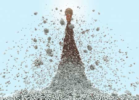 |
| A visualization of laser ablation depicts nanoparticle generation. (Image: Benjamin Hernandez, ORNL) |
| To corroborate their findings, the UVA scientists collaborated with a research group from the University of Duisburg-Essen, Germany. In 2018, their results were published in Nanoscale ("Two Mechanisms of Nanoparticle Generation in Picosecond Laser Ablation in Liquids: The Origin of the Bimodal Size Distribution"); the journal’s back cover featured a laser ablation image OLCF computer scientist Benjamin Hernandez created using SIGHT, a customizable visualization tool he developed. The OLCF is a US Department of Energy (DOE) Office of Science User Facility located at DOE’s Oak Ridge National Laboratory (ORNL). |
Following virtual clues |
| To differentiate between the sources of nanoparticles categorized as small (less than 10 nanometers) and large (10 or more nanometers), the team ran a series of molecular dynamics simulations on Titan, which modeled silver and gold targets in water irradiated by laser ablation. |
| “These metals are stable, inert, and do not actively react with the surrounding environment,” Zhigilei said. “Additionally, silver has useful antibacterial properties.” |
| The simulation results indicated that small nanoparticles are more likely to form from the condensation of metal vapor rapidly cooled through its interaction with water vapor, whereas large ones may emerge when hydrodynamic instabilities, which are unstable flows of one fluid through another fluid of a different density, cause the metal to disintegrate. |
| During ablation, laser pulses superheat part of the metal target’s surface, leading to an explosive decomposition of that region into a mixture of vapor and small liquid droplets. This hot mixture is then ejected from the irradiated target, forming the so-called ablation plume. Known as phase explosion or “explosive boiling,” this phenomenon has been studied extensively for laser ablation in a vacuum. |
| However, when ablation takes place in a liquid environment, the interaction of the ablation plume with the surrounding water complicates the process by slowing down the ablation plume, which leads to the formation of a hot metal layer pushing against the water. |
| This dynamic interaction can trigger a rapid succession of hydrodynamic instabilities in the molten metal layer, causing it to partially or entirely disintegrate and produce large nanoparticles. A well-known novelty item illustrates this behavior. |
| The team observed the movements of individual atoms to extrapolate useful information concerning both paths to nanoparticle generation. |
| “We had to quickly pivot from atoms on the scale of less than one nanometer to hundreds of nanometers, which required solving equations for hundreds of millions of atoms in our simulations,” Zhigilei said. “This type of work is only possible on large supercomputers like Titan.” |
| Both processes that lead to nanoparticle generation take place within a transient “reaction chamber” known as the cavitation bubble, which results from the interaction between the hot ablation plume and the liquid environment. By studying the bubble’s lifetime from start to finish, scientists can identify which types of nanoparticles emerge at certain stages. |
| “Irradiating a metal target in water with laser pulses creates a hot environment that leads to the formation, expansion, and collapse of a large bubble similar to those created by conventional boiling,” Zhigilei said. “Any nanoparticle generation process happens either within the bubble or in the interface between the ablation plume and the surface of the bubble.” |
| Complementary imaging experiments performed at the Center for Nanointegration Duisburg-Essen (CENIDE) confirmed the team’s computational findings by revealing the existence of smaller microbubbles containing nanoparticles that formed around the main cavitation bubble. |
| The CENIDE researchers also made videos demonstrating the production of gold nanoparticles and displaying a gold target immersed in a liquid ablation chamber. |
| "Naked" nanoparticles for medical application and catalyst surfaces which require high purity: Luckily, the nanoparticles made physically by laser ablation are free of residual chemicals (free of ligands) that may cause unwanted cross-effects or blocking of the surface. |
| Large-scale nanoparticle synthesis with a 500 Watt picosecond-pulsed laser system employing a polygon scanner reaching ultrasconic speed (up to 500 m/s) is shown in real time. This side view on the ablation chamber shows a gold target on the bottom of the water-filled vessel. The beautiful red color is caused by the so-called plasmon resonance of gold nanoparticles that are generated from the gold target, and documents the nanoparticle´s stability by eletrostatic repulsion. |
A blueprint for improvements |
| Scientists traditionally have relied on synthesis techniques to efficiently produce nanoparticles through a sequence of chemical reactions. Although this process allows for precise control over nanoparticle size, chemical contamination can prevent the resulting materials from functioning properly. Laser ablation avoids this pitfall by generating superior, clean nanoparticles while subtly molding metal into more suitable configurations. |
| “Laser ablation creates a completely clean colloidal solution of nanoparticles without using any other chemicals, and these pristine materials are ideal for biomedical applications,” Zhigilei said. “The results of our calculations may help to scale up this process and improve productivity so that ablation can eventually compete with chemical synthesis in terms of the number of nanoparticles produced.” |
| Finding the source of the size discrepancy paves the way to a future where researchers can optimize laser ablation to control the size of clean nanoparticles, making them cheaper and more readily available for potential biomedical purposes such as selectively killing cancer cells. |
| This achievement also exemplifies the benefits of laser technology while taking steps toward uncovering the fundamental factors that influence the outcomes of interactions between a laser pulse and a metal. This knowledge could lead to great strides in the team’s nanoparticle research, as well as advances in laser ablation and related techniques, which in turn would enable more precise interpretation of existing data. |
| Cheng-Yu Shih, lead author of the Nanoscale paper and a recent graduate of UVA, now works to combine modeling with experimental studies to further explore how different metals generate nanoparticles in response to laser ablation. |
| Zhigilei hopes the research will result in a breakthrough that removes the need for the tedious task of sorting small and large nanoparticles. |
| Source: Oak Ridge National Laboratory |
|
Subscribe to a free copy of one of our daily Nanowerk Newsletter Email Digests with a compilation of all of the day's news. |

