| Posted: Nov 15, 2017 |
Combining nanofluidics and machine learning to diagnose cancer(Nanowerk News) Researchers have developed an approach for exosome isolation, wherein millions of nanofluidic exosome sorting components are incorporated onto a single chip and work in parallel to isolate exosomes from clinical samples. |
| Nanoscale exosomes (30-200 nm diameter) released during fusion of the multivesicular endosomes with the plasma membrane, and which are found circulating in the blood, have been discovered to contain molecular information on their cells of origin, relevant for disease diagnostics, disease monitoring, and drug efficacy screening. |
| Reporting their findings in ACS Nano ("Combining Machine Learning and Nanofluidic Technology To Diagnose Pancreatic Cancer Using Exosomes"), the scientists from the University of Pennsylvania describe their approach to isolate specific subtypes of exosomes with robust, high throughput nanofluidics sorting. |
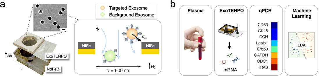 |
| ExoTENPO-based exosome capture. (a) A photograph of the ExoTENPO and the NdFeB external magnet with an SEM image of the magnetic nanopores; scale bar: 600 nm. (b) A schematic of the chip-based assay. From plasma, magnetically labeled exosomes are isolated, their mRNAs are isolated and profiled using qPCR, and the signature is found using a machine learning algorithm. (© ACS) (click on image to enlarge) |
| The researchers used linear discriminant analysis (LDA) on the exosomal RNA extracted from these exosomes to diagnose cancer in both a mouse model and human subjects and to diagnose precancerous lesions in a mouse model, all in blinded studies. |
| The team's exosome track-etched magnetic nanopore (ExoTENPO) chip was able to properly classify all patient samples and distinguish between healthy subjects and those with pancreatic cancer. |
| The detection of precancerous lesions in the murine model demonstrates the potential of using the ExoTENPO to screen for early stage pancreatic cancer. |
| LDA was used in this study because it is a relatively simple and well understood technique that can discriminate categorical variables using a set of continuous variables. |
| The scientists point out that more complicated machine learning algorithms can be used to further improve performance but were not explored in this study due to the limited number of samples. |
| According to the team, there are several aspects of the ExoTENPO that can be further developed to expand the system’s functionality to detect a wider range of diseases. The size of the ExoTENPO used in this study (d = 600 nm) could be made smaller or larger to tailor it to alternative extracellular vesicle targets, such as microvesicles (from 200 nm to 1 µm) or oncosomes (∼1 µm). |
| "By integrating nanoscale nucleic acid detection downstream of the ExoTENPO in future work, we can create a compact, self-contained device for a point-of-care use," the authors conclude their report. "Moreover, although this paper has focused on diagnostics, exosomes have been demonstrated in recent years to play a role in intracellular communication. Our chip's capability to specifically isolate exosome subpopulations can be an important tool in the study of this exosome-mediated communication." |
 By
Michael
Berger
– Michael is author of four books by the Royal Society of Chemistry:
Nano-Society: Pushing the Boundaries of Technology (2009),
Nanotechnology: The Future is Tiny (2016),
Nanoengineering: The Skills and Tools Making Technology Invisible (2019), and
Waste not! How Nanotechnologies Can Increase Efficiencies Throughout Society (2025)
Copyright ©
Nanowerk LLC
By
Michael
Berger
– Michael is author of four books by the Royal Society of Chemistry:
Nano-Society: Pushing the Boundaries of Technology (2009),
Nanotechnology: The Future is Tiny (2016),
Nanoengineering: The Skills and Tools Making Technology Invisible (2019), and
Waste not! How Nanotechnologies Can Increase Efficiencies Throughout Society (2025)
Copyright ©
Nanowerk LLC
|
|
Subscribe to a free copy of one of our daily Nanowerk Newsletter Email Digests with a compilation of all of the day's news. |