| Aug 29, 2008 |
Nanotomography produces incredible high-resolution 3-D images of nanostructures
|
|
(Nanowerk News) X-rays from a synchrotron can produce incredible high-resolution three-dimensional images of nanoscale crystal structures.
|
|
Researchers have combined medical imaging equipment with radiation from a particle accelerator to produce a powerful new method for three-dimensional (3D) imaging. The technique, developed by YU Shuhong, TIAN Yangchao and co-workers at the University of Science and Technology of China in Hefei,CAS, has already produced remarkable 3D renderings of a copper sulphide crystal, and could find many applications in materials development and the life sciences.
|
 |
| Schematic representation of moving filaments that serve as nanoshuttles, transporting and delivering cargo in a controlled manner. (Image: Diez group, Max Planck Institute of Molecular Cell Biology and Genetics)
|
|
Electron microscopes can provide detailed two-dimensional information on functional nanostructures, but are limited in applications involving complex 3D shapes. On the other hand, X-rays can penetrate to great depths and are used in medical computed-tomography scans to take images in slices that are then compiled to render a 3D image.
|
|
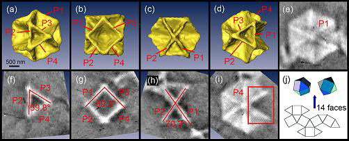
Yu, Tian and co-workers used the beamline from the National Synchrotron Radiation Laboratory in Hefei to provide a very powerful X-ray flux. The beam was carefully manipulated to make 3D images of a recently discovered type of copper sulphide crystal.
|
|
The images show that the crystal is made up of four hexagonal plates about 200 nanometres thick (pictured above). Together they form a cuboctahedron - a polyhedron with six square and eight triangular faces.
|
|
The image details indicate that the X-ray technique is useful to image nanomaterials with complex structures.
|
|
|

