| Jan 29, 2013 |
Quadruplex DNA found in human cells
|
|
(Nanowerk News) 60 years after Watson and Crick's ground breaking paper described the double helix structure of DNA, researchers at the University of Cambridge have observed four-stranded DNA structures within human cells. The discovery could open the door to novel cancer therapies and a new era for personalised medicine.
|
|
The findings mark the culmination of a 10 year investigation, part-funded by BBSRC, into the role of G-quadruplexes within the human genome. Working at the interface of chemistry and biology, from the hypothetical, through computational modelling to synthetic lab experiments, Professor Shankar Balasubramanian and Professor Steve Jackson's research groups have shown that G-quaruplexes, so-called because they form in guanine-rich DNA sequences, are not just a structural curiosity - they form at sites across the genome in living cells.
|
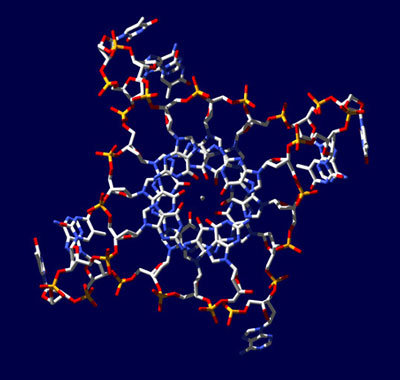 |
| X-ray crystal structure of a G-quadruplex formed from the DNA sequence found in human genomes.
|
|
"We've come such a long way from thinking that we understand the genome - and it appeared that this structure could tell us something new," says Balasubramanian, whose co-invention of Solexa Sequencing, an ultrafast way to sequence DNA, led to his award as BBSRC Innovator of the Year in 2010.
|
|
But it wasn't until a strong association was noticed with genes responsible for cell proliferation that Balasubramanian and others began to suspect that G-quadruplexes might be a potential target for cancer therapy. In 2012, the Cambridge team showed for the very first time that these structures form in cancer cells. Then, using a small synthetic drug called pyridostatin, developed by Senior Research Associate Dr Raphaël Rodriguez, to interact with these complex structures they were able to prevent proliferation of cancer cells. This work was published in Nature Chemical Biology ("Small-molecule–induced DNA damage identifies alternative DNA structures in human genes").
|
|
Cell cycle clues
|
|
In their latest study, funded by Cancer Research UK, PhD student Giulia Biffi and the team have identified G-quadruplexes in cancer cells using fluorescent biomarkers, which allowed them to pinpoint the time and place of the structures' emergence within the cell cycle. The paper was published last week in Nature Chemistry ("Quantitative visualization of DNA G-quadruplex structures in human cells"). The research goes on to show clear links between concentrations of four-stranded quadruplexes and the process of DNA replication, which is pivotal to cell division and production.
|
|
While quadruplex DNA is found fairly consistently throughout the genome of human cells and their division cycles, a marked increase was shown when the fluorescent staining grew more intense during the 's-phase' - the point in a cell cycle where DNA replicates before the cell divides.
|
|
Cancers are usually driven by genes called oncogenes that have mutated to increase DNA replication - causing cell proliferation to spiral out of control, and leading to tumour growth. The increased DNA replication rate during oncogenesis leads to an intensity in the quadruplex structures.
|
|
Using Solexa sequencing to look across the whole genome, the collaboration had previously identified that SRC, a major gene associated with breast cancer, is one of several hotspots on the genome targeted by pyridostatin.
|
|
"The data supports the idea that certain cancer genes can be usefully interfered with by small molecules designed to bind specific DNA sequences," said Balasubramanian.
|
|
"Many current cancer treatments attack DNA, but it's not clear what the rules are. We don't even know where in the genome some of them react - it can be a scattergun approach. The possibility that particular cancer cells harbouring genes with these motifs can now be targeted, and appear to be more vulnerable to interference than normal cells, is a thrilling prospect.
|

