| Aug 30, 2013 |
Membranes contain beautiful patterns - but their function is a mystery
|
|
(Nanowerk News) Biological cells are surrounded by a membrane, and here some of the most important processes for sustaining life take place. There can also be something very beautiful happening in membranes, researchers from the University of Southern Denmark have discovered: Membranes can contain beautiful, mysterious patterns.
|
|
"We do not yet know what the possible biological function of this might be. There should be a reason for the patterns, we just have not discovered it yet", says associate professor Adam Cohen Simonsen, Department of Physics, Chemistry and Pharmacy, University of Southern Denmark.
|
|
With his colleagues Jes Dreier, Jonathan Brewer, John Hjort Ipsen and Uffe Bernchou (now Odense University Hospital) from the research group MEMPHYS at Department of Physics, Chemistry and Pharmacy at the University of Southern Denmark, he has discovered that cell membranes can form spectacular patterns ("Hydrophobic Mismatch Triggering Texture Defects in Membrane Gel Domains").
|
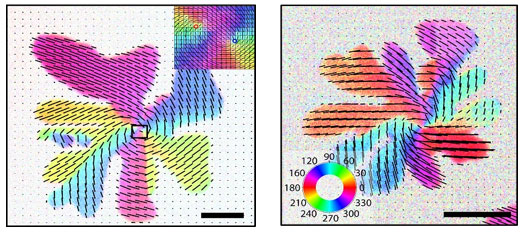 |
| Examples of spiral patterns. The colors indicate the direction of the lipids in the membrane, as shown in the color code that is inserted in image top right. The figure inserted in the image top left shows an enlargement of the pattern of the black square. The length of the black line is 5 micrometers.
|
|
The patterns are formed by highly organized lipids (fats) which make up the membrane, and the patterns vary according to conditions such as temperature and the particular type of lipid molecules. The scientists have recently published their latest work identifying two different patterns: a spiral pattern and a uniform pattern.
|
|
"This is just the beginning. There may be more than the two patterns we have identified", says Adam Cohen Simonsen.
|
|
The patterns are so difficult to "see" that only few research groups in the world yet have the experimental facilities required to visualize the mysterious patterns. In the laboratory at the University of Southern Denmark in Odense the scientists have constructed a custom-designed light microscope, which takes advantage of polarized light to reveal the patterns, and the scientists have managed to produce detailed microscope images of them.
|
|
The phenomenon - that membranes can form such so-called texture patterns - was discovered at SDU five years ago. Now the researchers have discovered the possibility of more than one pattern and, using artificial membranes, they have identified the conditions under which two particular types of patterns are formed in the laboratory.
|
|
Artificial membranes have the advantage that they can easily be spread out on a flat surface and placed under a microscope, while a cell membrane taken from a biological organism is round and much more difficult to work with. Like the membranes of living cells the artificial laboratory membranes are composed of different molecules, in this case, a particular family of lipid molecules – phospholipids. These are also found in the biological cell membranes.
|
|
"The precise type of lipid molecules, that the membrane consists of, appears to play a big role. Some lipid molecules produce spiral patterns and other lipid molecules produce the uniform pattern. The molecules we use are not all of the same length, and therefore different molecules create different membrane thicknesses. The thickness of the membrane can vary from say 5 nm to 6 nm, depending on the molecules used", Adam Cohen Simonsen explains.
|
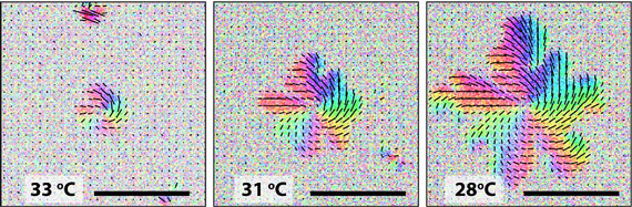 |
| A series of images, which illustrates the formation of a spiral pattern over a period during which the temperature decreases. First at app. 33 degrees a germ is created. From this the spiral grows and it ends with a spiral pattern at 28 degrees. This takes approx. five minutes. The length of the black line is 5 micrometers.
|
|
When he speaks about the thickness of the membrane, the important quantity is in fact the change in thickness of an elevated area of the membrane, where the lipid molecules are more densely packed. Such a densely packed region of the membrane is called a membrane domain, and it is in such areas that the patterns exist.
|
|
"The first pattern we discovered was the spiral pattern. It can be compared to the pattern that is created when you rake long grass in patterns. Lipid molecules are in this picture, like the leaves of grass, organized in a distinctive direction. The next pattern observed was the uniform pattern. Here we saw how lipid molecules organized themselves in a much simpler way, with all molecules pointing in the same direction. Our results suggest that it is the change in membrane thickness across a domain that determines whether one or the other pattern is formed", says Adam Cohen Simonsen.
|
|
It takes a few minutes for a spiral pattern to form in the laboratory where the growth takes place under controlled conditions. The formation starts at a relatively high temperature, 55 degrees Celsius, where the lipid molecules are in a liquid state and slide effortlessly around between each other. When the temperature drops, the lipid molecules start condensing together, and at app. 34 degrees the pattern formation starts.
|
|
"When we studied the spiral pattern, we saw that a nucleus was formed at 34 degrees, and that the spiral domain grew from the nucleus. It took approximately five minutes for it to finish", says Adam Cohen Simonsen.
|
|
"We have now shown that the type of pattern depends on which kind of lipid molecules that is present in the cell membrane. But what the function is – or what significance, these patterns may have for the biological functionality of living cells – that is something we do not know yet. It is the next big question", Adam Cohen Simonsen concludes.
|

