| Aug 14, 2014 |
Researchers identify key mechanism that drives movement in living cells
|
|
(Nanowerk News) A team of international researchers led by Professor Lim Chwee Teck, Principal Investigator, and Dr Hiroaki Hirata, Research Fellow, at the Mechanobiology Institute at the National University of Singapore (NUS), together with Professor Masahiro Sokabe from Nagoya University Graduate School of Medicine, has recently discovered that living cell migration is regulated by the engagement of a force transmitter composed of vinculin and talin, two types of cytoskeletal protein. The researchers showed that force-dependent vinculin binding to talin plays a critical role in mechanically connecting the actin cytoskeleton to the extracellular substrate to contribute towards cell migration.
|
|
This understanding of the fundamental machinery that drives living cells movement is crucial in paving the way for physiological and pathological processes in modern medicine, ranging from normal tissue development to treating cardiovascular disorders and cancer metastasis. The findings were recently published in an international journal, American Journal of Physiology - Cell Physiology ("Force-dependent vinculin binding to talin in live cells: a crucial step in anchoring the actin cytoskeleton to focal adhesions").
|
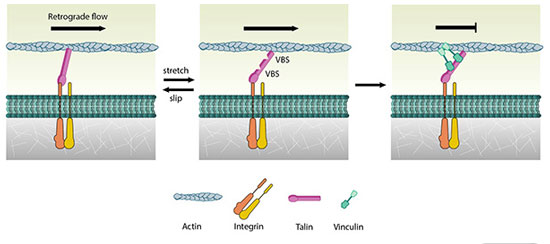 |
| Schematic showing the model of vinculin binding to the integrin-talin-actin complex, subsequently arresting the actin filament retrograde flow.
|
|
Demystifying cell movement
|
|
Individual cells possess distinct structures, known as focal adhesions, which allow them to grip onto a surface of the extracellular substrate. Inside the cell, focal adhesions are physically linked to a network of filaments, composed of a protein called actin, that form near the protruding cell membrane. These are highly dynamic and are constantly undergoing assembly and disassembly. Due to their dynamic nature, as well as the activity of protein known as myosin, the actin filaments are constantly moving inwards, towards the center of the cell.
|
|
For cells to move forward, cells have to transmit force from dynamic actin filaments to the extracellular substrate through focal adhesions. The force transmitter is engaged when force is exerted onto it. This is akin to the “automatic clutches” in vehicles. The researchers found that the force-dependent vinculin binding to talin underlies engagement of the transmitter. When the transmitter was artificially disengaged by inhibiting the vinculin-talin binding, the cells failed to move as they no longer transmit force.
|
|
It was observed that talin solely serves as slipping connection between the moving actin filament and the adhesion molecule called integrin. However, force-dependent vinculin binding to talin forms a stable integrin-talin-vinculin-actin link. The link with multiple vinculin molecules could sustain a far greater force, and this was sufficient to tether actin filaments to focal adhesions.
|
|
Although the actin filaments no longer move inwards, they are still growing near the cell membrane. This enables the membrane to protrude outwards around the growing filaments. Eventually, new adhesions form at the tip of the protrusion, allowing the cell to grab onto the substrate surface. These cycles allow the cell to move forward.
|
|
With better understanding of these processes, the researchers hope to identify new technologies and therapies targeted at encouraging or preventing cell motility.
|
|
The team has also developed a dominant-negative form of vinculin which inhibits the vinculin binding to talin, thereby inhibiting engagement of the force transmitter. If a method can be developed to introduce this dominant negative mutant specifically into cancer cells, invasion and metastasis of cancer cells could largely be suppressed.
|

