| Jun 24, 2019 |
Helping the body's ability to grow bone
|
|
(Nanowerk News) For the first time, scientists have been able to study how well synthetic bone grafts stand up to the rigors and 'strains' of life, and how quickly they help bone re-grow and repair.
|
|
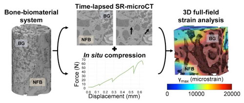
Researchers led by Dr Gianluca Tozzi, at the University of Portsmouth, are the first to examine the strains between bone and graft from animal models in 3D and in microscopic detail.
|
 |
| This is a graphical abstract summarizing the experiment. (Image: University of Portsmouth) (click on image to enlarge)
|
|
Dr Tozzi hopes this window on to living bone grafts will help scientists find ways to improve the body's ability to regrow its own bone, and more chance surgeons can predict the success of a synthetic graft.
|
|
He said: "Every three seconds a person breaks a bone due to increased bone fragility. Fragile bones break easily and are also more difficult to repair, particularly when the defect area is extended. It's vital we understand what is happening where bone meets graft so we can better engineer sophisticated replacement materials.
|
|
"Bones are very complex biological tissues and a synthetic bone substitute needs to have specific requirements to allow blood supply and encourage new bone growth.
|
|
"In this sense, the new generation of synthetic grafts have the potential to be resorbed by the body in time, allowing gradual bone regeneration in the defect site. However, biomaterials that degrade too quickly don't allow enough time for the new bone to grow, and grafts that degrade too slowly can cause mechanical instability to the implantation site. It's important to get it right."
|
|
Millions of people a year in the UK are given a bone graft. They're commonly used in the spine, hip, knee and ankle. Their role is bridge gaps in a broken bone that are too large for the bone to close on its own. They're also used in dental implants, to help teeth attach to the jawbone.
|
|
Some grafts can be made using a fragment of the patient's own bone or other sources, but this is more invasive and can cause adverse reactions. Therefore, it's becoming increasingly common for grafts to be made by synthetic materials, including glass, ceramics and even, in very small joints, plaster of Paris.
|
|
Dr Tozzi and colleagues have been using synchrotron X-ray computed tomography (SR-XCT) at the Diamond Light Source and in lab-based systems at the Zeiss Global Centre at the University of Portsmouth to better understand the performance of graft materials and their ability to promote bone healing.
|
|
In a recently published study in ACS Biomaterials Science & Engineering("Full-Field Strain Analysis of Bone–Biomaterial Systems Produced by the Implantation of Osteoregenerative Biomaterials in an Ovine Model"), they examined the micromechanics and microdamage evolution of four different bone-biomaterial systems combining high-resolution synchrotron tomography, in situ mechanics and digital volume correlation.
|
|
Dr Tozzi said: "It's essential we are able to look at the interface between bone and graft and judge their load-bearing capability in order to understand both biological integration and structural integrity of the intervention.
|
|
"By carrying out time-lapse experiments of such constructs, we could observe damage progression and see, for the first time, how strain could be used to understand and potentially predict clinical outcome of biomaterials in a living body, improving our knowledge significantly."
|


