| Aug 28, 2019 |
Retina-on-a-chip provides powerful tool for studying eye disease
|
|
(Nanowerk News) The development of a retina-on-a-chip, which combines living human cells with an artificial tissue-like system, has been described in the open-access journal eLife ("Merging organoid and organ-on-a-chip technology to generate complex multi-layer tissue models in a human Retina-on-a-Chip platform").
|
|
This cutting-edge tool may provide a useful alternative to existing models for studying eye disease and allow scientists to test the effects of drugs on the retina more efficiently.
|
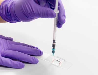 |
| Organ-on-a-chip system. (Image: Fraunhofer Institute for Interfacial Engineering and Biotechnology)
|
|
Many diseases that cause blindness harm the retina, a thin layer of tissue at the back of the eye that helps collect light and relay visual information to the brain. The retina is also vulnerable to harmful side effects of drugs used to treat other diseases such as cancer.
|
|
Currently, scientists often rely on animals or retina organoids, tiny retina-like structures grown from human stem cells, to study eye diseases and drug side effects. But results from studies in both models often fail to describe disease and drug effects in people accurately. As a result, a team of scientists have tried to recreate a retina for testing purposes using engineering techniques.
|
|
“It is extremely challenging, if not almost impossible, to recapitulate the complex tissue architecture of the human retina solely using engineering approaches,” explains Christopher Probst, Postdoctoral Researcher at the Fraunhofer Institute for Interfacial Engineering and Biotechnology in Stuttgart, Germany, and co-lead author of the current study.
|
|
To overcome these challenges, the scientists coaxed human pluripotent stem cells to develop into several different types of retina cells on artificial tissue. This tissue recreates the environment that cells would experience in the body and delivers nutrients and drugs to the cells through a system that mimics human blood vessels.
|
|
“This combination of approaches enabled us to successfully create a complex multi-layer structure that includes all cell types and layers present in retinal organoids, connected to a retinal pigment epithelium layer,” says co-lead author Kevin Achberger, Postdoctoral Researcher at the Department of Neuroanatomy & Developmental Biology at the Eberhard Karls University of Tübingen, Germany. “It is the first demonstration of a 3D retinal model that recreates many of the structural characteristics of the human retina and behaves in a similar way.”
|
|
The team treated their retina-on-the-chip with the anti-malaria drug chloroquine and the antibiotic gentamicin, which are toxic to the retina. They found that the drugs had a toxic effect on the retinal cells in the model, suggesting that it could be a useful tool for testing for harmful drug effects.
|
|
“One advantage of this tiny model is that it could be used as part of an automated system to test hundreds of drugs for harmful effects on the retina very quickly,” Achberger says. “Also, it may enable scientists to take stem cells from a specific patient and study both the disease and potential treatments in that individual’s own cells.”
|
|
“This new approach combines two promising technologies – organoids and organ-on-a-chip – and has the potential to revolutionise drug development and usher in a new era of personalised medicine,” concludes senior author Peter Loskill, Assistant Professor for Experimental Regenerative Medicine at the Eberhard Karls University of Tübingen, and head of the Fraunhofer Attract group Organ-on-a-Chip at the Fraunhofer Institute for Interfacial Engineering and Biotechnology. His laboratory, which spans the two institutes, is already developing similar organ-on-a-chip technology for the heart, fat, pancreas and more.
|

