| Sep 05, 2019 |
New method for imaging biological molecules
|
|
(Nanowerk News) Researchers at Karolinska Institutet in Sweden have, together with colleagues from Aalto University in Finland, developed a new method for creating images of molecules in cells or tissue samples. The method is based on the use of DNA snippets and is called DNA microscopy. The approach is currently described in the scientific journal PNAS ("A computational framework for DNA sequencing microscopy").
|
|
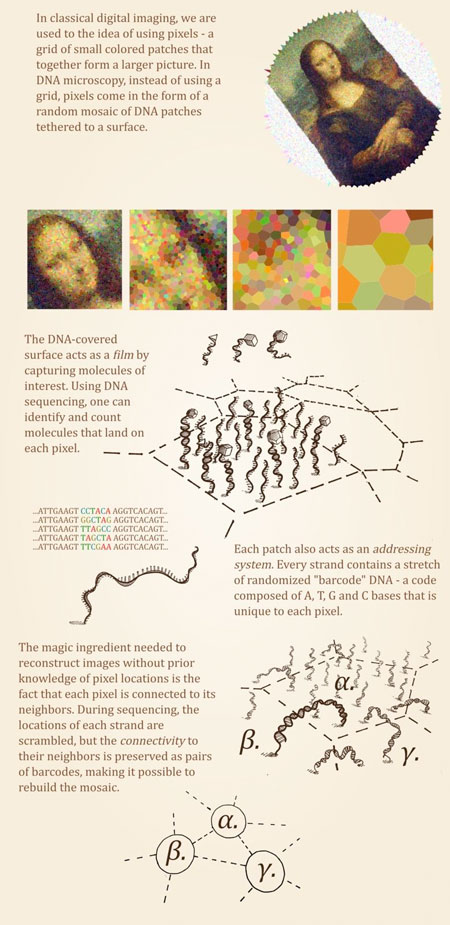
The new method involves researchers in the first stage mixing up a cell or tissue sample with short sequences of single-stranded DNA, selected to attach to the specific molecules that are going to be studied. If it for example involves a specific protein that is going to be investigated, small DNA snippets are used that bind to this particular protein. In the next stage, enzymes are fed to the short DNA sequences to connect and form DNA molecules.
|
|
By analysing these newly formed DNA molecules with so-called DNA sequencing, it is possible to see exactly which DNA snippets have ended up next to each other. Based on this information, you can add a puzzle that shows how all of the DNA sequences must be connected.
|
|
Since DNA sequences are attached to the molecules that are being represented, it is possible to understand how abundant they occur and where they are in the cells. What researchers are now publishing is a mathematical model that makes it possible to calculate this as well as create images from such information.
|
 |
| (Illustration: Ian Hoffecker)
|
|
"You can liken it to the table placement game at a wedding where each guest gets a note that matches the person next to them at the table. If you have all of these notes you can recreate the table placement. In our experiments, the notes represent the DNA-snippets and the guests are the molecules that the snippets are attached to," says Björn Högberg, professor at the Department of Medical Biochemistry and Biophysics at Karolinska Institutet.
|
|
The advantage of this method, which scientists call DNA microscopy, is that it makes it possible to search for specific molecules within a larger material, such as a whole cell collection or a tissue sample. With traditional microscopy, where you have to look at one area at a time, it is very time-consuming. But with DNA microscopy it is possible to, for example, screen for certain molecules and examine how frequent they are.
|
|
The method also makes it possible to see what role the immediate environment plays in the life of a cell, i.e. how micro-environment might affect possible disease development - to mention only two of several possible useful areas when it comes to DNA microscopy.
|
|
"This is a tool that can be used to gain a better understanding of how biology works and how cells work together. This knowledge can provide us with a better picture of how different diseases develop. In the long run, this tool can also provide opportunities for safer diagnostics," says Ian Hoffecker, researcher in Björn Högberg's Research Group at the Department of Medical Biochemistry and Biophysics at Karolinska Institutet and the coordinator of the study.
|

