| Aug 27, 2022 |
Scientists grow 'synthetic' embryo with brain and beating heart from stem cells
|
|
(Nanowerk News) Researchers from the University of Cambridge have created model embryos from mouse stem cells that form a brain, a beating heart, and the foundations of all the other organs of the body – a new avenue for recreating the first stages of life.
|
|
The team, led by Professor Magdalena Zernicka-Goetz, developed the embryo model without eggs or sperm, and instead used stem cells – the body’s master cells, which can develop into almost any cell type in the body.
|
|
The researchers mimicked natural processes in the lab by guiding the three types of stem cells found in early mammalian development to the point where they start interacting. By inducing the expression of a particular set of genes and establishing a unique environment for their interactions, the researchers were able to get the stem cells to ‘talk’ to each other.
|
|
The stem cells self-organised into structures that progressed through the successive developmental stages until they had beating hearts and the foundations of the brain, as well as the yolk sac where the embryo develops and gets nutrients from in its first weeks. Unlike other synthetic embryos, the Cambridge-developed models reached the point where the entire brain, including the anterior portion, began to develop.
|
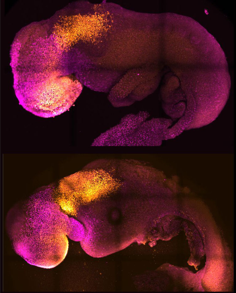 |
| Natural (top) and synthetic (bottom) embryos side by side to show comparable brain and heart formation. (Image: Amadei and Handford)
|
|
The team say their results, the result of more than a decade of research that progressively led to more and more complex embryo-like structures and reported in the journal Nature ("Synthetic embryos complete gastrulation to neurulation and organogenesis"), could help researchers understand why some embryos fail while others go on to develop into a healthy pregnancy. Additionally, the results could be used to guide repair and development of synthetic human organs for transplantation.
|
|
“Our mouse embryo model not only develops a brain, but also a beating heart, all the components that go on to make up the body,” said Zernicka-Goetz, Professor in Mammalian Development and Stem Cell Biology in Cambridge’s Department of Physiology, Development and Neuroscience, adding: “It’s just unbelievable that we’ve got this far. This has been the dream of our community for years, and a major focus of our work for a decade, and finally we’ve done it.”
|
|
For a human embryo to develop successfully, there needs to be a ‘dialogue’ between the tissues that will become the embryo, and the tissues that will connect the embryo to the mother. In the first week after fertilisation, three types of stem cells develop: one will eventually become the tissues of the body, and the other two support the embryo’s development. One of these extraembryonic stem cell types will become the placenta, which connects the foetus to the mother and provides oxygen and nutrients; and the second is the yolk sac, where the embryo grows and where it gets its nutrients from in early development.
|
|
Many pregnancies fail at the point when the three types of stem cells begin to send mechanical and chemical signals to each other, which tell the embryo how to develop properly.
|
|
“So many pregnancies fail around this time, before most women realise they are pregnant,” said Zernicka-Goetz, who is also Professor of Biology and Biological Engineering at Caltech. “This period is the foundation for everything else that follows in pregnancy. If it goes wrong, the pregnancy will fail.”
|
|
Over the past decade, Professor Zernicka-Goetz’s group in Cambridge has been studying these earliest stages of pregnancy, in order to understand why some pregnancies fail and some succeed. |
|
“The stem cell embryo model is important because it gives us accessibility to the developing structure at a stage that is normally hidden from us due to the implantation of the tiny embryo into the mother’s womb."
|
|
Zernicka-Goetz added: “This accessibility allows us to manipulate genes to understand their developmental roles in a model experimental system.”
|
|
To guide the development of their synthetic embryo, the researchers put together cultured stem cells representing each of the three types of tissue in the right proportions and environment to promote their growth and communication with each other, eventually self-assembling into an embryo.
|
|
The researchers found that the extraembryonic cells signal to embryonic cells by chemical signals but also mechanistically, or through touch, guiding the embryo’s development.
|
|
“This period of human life is so mysterious, so to be able to see how it happens in a dish – to have access to these individual stem cells, to understand why so many pregnancies fail and how we might be able to prevent that from happening – is quite special,” said Zernicka-Goetz. “We looked at the dialogue that has to happen between the different types of stem cell at that time – we’ve shown how it occurs and how it can go wrong.”
|
|
A major advance in the study is the ability to generate the entire brain, in particular the anterior part, which has been a major goal in the development of synthetic embryos.
|
|
This works in Zernicka-Goetz’s system because this part of the brain requires signals from one of the extraembryonic tissues to be able to develop. The team thought that this might be taking place from their 2018 and 2021 studies, which used the same component cells to develop into embryos at a slightly earlier stage. Now, by pushing development just one day further, they can definitively say that their model is the very first to signal development of the anterior, and in fact the whole, brain.
|
|
“This opens new possibilities to study the mechanisms of neurodevelopment in an experimental model,” said Zernicka-Goetz. “In fact, we demonstrate the proof of this principle in the paper by knocking out a gene already known to be essential for formation of the neural tube, precursor of the nervous system, and for brain and eye development. In the absence of this gene, the synthetic embryos show exactly the known defects in brain development as in an animal carrying this mutation. This means we can begin to apply this kind of approach to the many genes with unknown function in brain development.”
|
|
While the current research was carried out in mouse models, the researchers are developing similar human models with the potential to be directed towards the generation of specific organ types - to understand mechanisms behind crucial processes that would be otherwise impossible to study in real embryos. At present, UK law permits human embryos to be studied in the laboratory only up to the 14th day of development.
|
|
If the methods developed by Zernicka-Goetz’s team are shown to be successful with human stem cells in future, they could also be used to guide development of synthetic organs for patients awaiting transplants.
|
|
“There are so many people around the world who wait for years for organ transplants,” said Zernicka-Goetz. “What makes our work so exciting is that the knowledge coming out of it could be used to grow correct synthetic human organs to save lives that are currently lost. It should also be possible to affect and heal adult organs by using the knowledge we have on how they are made.
|

