| Mar 28, 2019 |
3D-printed tissues may keep athletes in action
|
|
(Nanowerk News) Bioscientists are moving closer to 3D-printed artificial tissues to help heal bone and cartilage typically damaged in sports-related injuries to knees, ankles and elbows.
|
|
Scientists at Rice University and the University of Maryland reported their first success at engineering scaffolds that replicate the physical characteristics of osteochondral tissue – basically, hard bone beneath a compressible layer of cartilage that appears as the smooth surface on the ends of long bones.
|
|
Injuries to these bones, from small cracks to pieces that break off, can be painful and often stop athletes’ careers in their tracks. Osteochondral injuries can also lead to disabling arthritis.
|
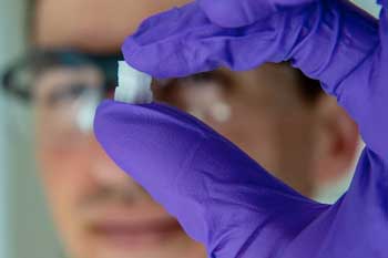 |
| Rice University graduate student Sean Bittner holds a 3D-printed scaffold created to help heal osteochondral injuries. The initial study is a proof-of-concept to see if printed structures can mimic the gradual transition from smooth, compressible cartilage to hard bone at the end of long bones. (Image: Jeff Fitlow/Rice University)
|
|
The gradient nature of cartilage-into-bone and its porosity have made it difficult to reproduce in the lab, but Rice scientists led by bioengineer Antonios Mikos and graduate student Sean Bittner have used 3D printing to fabricate what they believe will eventually be a suitable material for implantation.
|
|
Their results are reported in Acta Biomaterialia ("Fabrication and mechanical characterization of 3D printed vertical uniform and gradient scaffolds for bone and osteochondral tissue engineering").
|
|
“Athletes are disproportionately affected by these injuries, but they can affect everybody,” said Bittner, a third-year bioengineering graduate student at Rice, a National Science Foundation fellow and lead author of the paper. “I think this will be a powerful tool to help people with common sports injuries.”
|
|
The key is mimicking tissue that turns gradually from cartilage (chondral tissue) at the surface to bone (osteo) underneath. The Biomaterials Lab at Rice printed a scaffold with custom mixtures of a polymer for the former and a ceramic for the latter with imbedded pores that would allow the patient’s own cells and blood vessels to infiltrate the implant, eventually allowing it to become part of the natural bone and cartilage.
|
|
“For the most part, the composition will be the same from patient to patient,” Bittner said. “There’s porosity included so vasculature can grow in from the native bone. We don’t have to fabricate the blood vessels ourselves.”
|
|
The future of the project will involve figuring out how to print an osteochondral implant that perfectly fits the patient and allows the porous implant to grow into and knit with the bone and cartilage.
|
|
Mikos said the collaboration is a great early success for the Center for Engineering Complex Tissues (CECT), a National Institutes of Health center at Maryland, Rice and the Wake Forest School of Medicine developing bioprinting tools to address basic scientific questions and translate new knowledge into clinical practice.
|
|
“In that context, what we’ve done here is impactful and may lead to new regenerative medicine solutions,” Mikos said.
|

