| Sep 09, 2020 |
Oxygen-releasing bioink for 3D bioprinting |
| (Nanowerk News) Engineering new tissues can be used to alleviate shortages of organs in transplantation, as well as to develop physiological models for drug discovery applications. One of the emerging approaches to building tissues is through 3D Printing, where cells and materials can be combined to make inks that can generate tissue structures. |
| One of the limitations for making new tissues is that they require oxygen to survive. This oxygen is delivered through blood vessels, which take a few days to develop in a transplanted tissue. |
| A collaborative team, which includes a group from the Terasaki Institute for Biomedical Innovation (TIBI), has developed a bioink that offers a solution to this problem. This bioink can generate oxygen and deliver it to cells in 3D printed tissues to keep them alive before blood vessels penetrate the tissue. Therefore, when it is used in 3D bioprinting to construct tissue implants, the ability of cells to regenerate new tissue is greatly enhanced. |
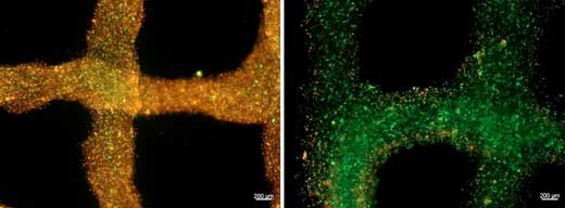 |
| Heart cells in a bioink (left) without oxygen support and (right) with oxygen-releasing capabilities. Live cells are stained green, dead cells in red. (Image: Khademhosseini Lab) |
| As discussed in their recent publication in Advanced Healthcare Materials ("3D Bioprinting of Oxygenated Cell-Laden Gelatin Methacryloyl Constructs"), the TIBI group tested their oxygen-generating bioink extensively to optimize its chemical and physical properties. |
| As a result, oxygen was delivered to the cells in the tissue constructs for the period necessary for blood vessels to develop fully. These blood vessels would then be able to resume their oxygen delivery functions, and the cells would have the additional support needed to grow and regenerate new tissue. |
| The group also conducted separate experiments on tissue constructs using two different types of cells, including cardiac cells and muscle cells, and they demonstrated the positive effects of using the new bioink. |
| "By delivering oxygen to the implanted cells, we would be able to improve the tissue functionality and integration to the host tissue. A similar approach can be used to make functional tissues with improved survival for drug screening applications and pathophysiological studies within a long period of time," said Samad Ahadian, Ph.D., lead investigator of the Terasaki Institute team. |
| Such developments can result in a variety of medical applications, including the enhancement of tissue regeneration in patients who have suffered a heart attack. These patients have experienced a period in which their heart is without a sufficient supply of oxygen. Therefore, there may be damage or a higher risk of harm to parts of their cardiac tissue. Tissue implants using the new bioink may help not only to increase the survival of the affected cardiac cells but can also help to support the growth and formation of the cardiac blood vessels. |
| Source: Terasaki Institute for Biomedical Innovation |

