| Sep 19, 2023 |
'Bioprinting' living brain cell networks in the lab
|
|
(Nanowerk News) Monash University Engineering researchers have successfully used “bioinks” containing living nerve cells (neurons) to print 3D nerve networks that can grow in the laboratory and transmit and respond to nerve signals.
|
|
The research has been published in Advanced Healthcare Materials ("3D Functional Neuronal Networks in Free-Standing Bioprinted Hydrogel Constructs").
|
|
Using a tissue engineering approach, and bioprinting with two bioinks containing living cells and non-cell materials respectively, the researchers were able to mimic the arrangement of grey matter and white matter seen in the brain.
|
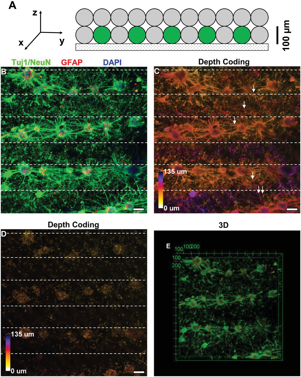 |
| Bioprinted cortical neurons and astrocytes after in vitro culture at 7DIV. A) Cross-section view of bioprinted structure consisting of cellular (green) and acellular (grey) strands. B) Maximum intensity projection of patterned structure: cellular–acellular–cellular. Neurons (NeuN, green), astrocytes (GFAP, red), and nuclei (DAPI, blue) were colocalized and developed complex structures. Axons (Tuj1, green) originating from the proximal cellular strands projected across the distal cellular strand. C) Depth-coding of Tuj1/NeuN showed the development of axonal projections across strands. White arrows indicate neurites that belong to different focal planes. D) Depth-coding of DAPI shows localization of cell nuclei. E) Confocal image in 3D. Depth-coding was represented using a color bar: red, closest to the glass substrate, and blue, 135 µm away from the substrate. Scale bar, 100 µm. (Image: Monash University)
|
|
Professor John Forsythe of the Department of Materials Science and Engineering, who is leading the research, said while two-dimensional nerve cell cultures have previously been used to study the formation of nerve networks and disease mechanisms, those relatively flat structures don’t reflect the way neurons grow and interact with their surroundings.
|
|
“The networks grown in this research closely replicated the 3D nature of circuits in a living brain, where nerve cells extend processes called neurites to form connections between different layers of the cortex,” said Professor Forsythe.
|
|
“We found that the projections growing from neurons in the printed ‘grey matter’ or cellular layer readily grew through the ‘white matter’ layer and used it as a ‘highway’ to communicate with neurons in other layers.
|
|
“Not only were we able to construct a basic layout similar to what we see in regions of the brain, we found that the neurons actually behaved and performed in a similar manner.”
|
|
Sensitive electrophysiological measurements confirmed spontaneous nerve-like activity taking place in the 3D neuronal networks in addition to responses evoked by electrical and drug stimulation.
|
|
The presence of detectable electrical activity in tissue engineered 3D networks represents a significant step forward in the field of neuroscience and bioprinting.
|
|
Bioprinted 3D neural networks are likely to be a promising platform for studying how nerves and nerve networks form and grow, investigating how some diseases affect neurotransmission, and screening drugs for their effects on nerve cells and the nervous system.
|

