| Feb 05, 2024 |
Watching the enzymes that convert plant fiber into simple sugars
|
|
(Nanowerk News) Research from Lawrence Berkeley National Laboratory (Berkeley Lab), Lawrence Livermore National Laboratory (LLNL), and UC Davis sheds new light on how to access the sugars locked up in plants to produce petroleum-free fuels, chemicals, and medicines.
|
|
Using microbes to convert grasses, weeds, wood, and other plant residues into sustainable products will be key to achieving carbon neutrality and could even help eliminate drug shortages. But cellulose, the tough tissue that makes up a large proportion of herbaceous and woody plant bodies, is hard to break down into its composite sugars, which the microbes need to build other molecules. Only organisms that have evolved specialized enzymes, or those that host microbiomes of those organisms, are able to get sugars from cellulose-rich plant matter.
|
|
Scientists are studying how these enzymes work so that they can develop more efficient methods to convert plant waste into sweet ingredients.
|
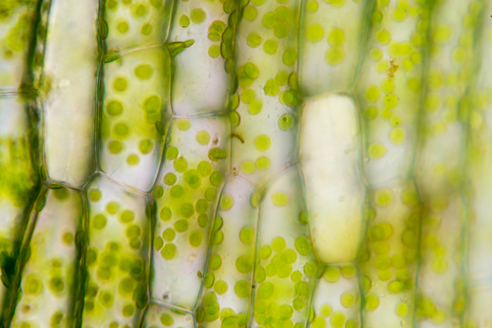 |
| A microscopy image of plant cells showing their rigid walls, which are given structure by cellulose. (Image: Rattiya Thongdumhyu / Shutterstock)
|
|
“We want to utilize plant residues and there is a lot of plant waste out there,” said project co-leader Tina Jeoh, a professor of biological and agricultural engineering at UC Davis. “These sugars are key to establishing a bioeconomy built on cycling renewable carbon for biofuel, biochemical, and biomaterial alternatives to fossil fuel-sourced versions.”
|
|
Jeoh and her colleagues used a technique developed at the Berkeley Synchrotron Infrared Structural Biology (BSISB) Imaging Program. The technique combines a novel microfluidic device and infrared spectroscopy to study how a cellulose-degrading enzyme works in real time.
|
|
Their work was recently published in the journal Green Chemistry ("Spatiotemporal dynamics of cellulose during enzymatic hydrolysis studied by infrared spectromicroscopy").
|
|
Cellulose is composed of many glucose molecules, each joined together by a single covalent bond. The long chains of glucose are twisted together into complex rope-like structures called fibrils, which stay in that formation thanks to many hydrogen bonds between the tightly-organized glucoses. Scientists have hypothesized that these hydrogen bonds are the reason why cellulose-chopping enzymes are so slow – they act as obstacles blocking access to the covalent bond.
|
|
To finally understand exactly what is happening during these reactions, a pair of Berkeley Lab researchers designed an experimental system that could provide information about how the atomic structure of the cellulose changed while the enzyme was working. The system consists of a small disk-like device that holds a tiny quantity of fluid containing cellulose, from green algae, and tiny amount of an enzyme derived from a fungi. The device moves the two fluids together, allowing the reaction to begin, in the path of a powerful beam of infrared light generated by the Berkeley Lab’s Advanced Light Source (ALS). Detectors near the device then measure how the light was absorbed by the combined fluids at different time intervals when exposed to the beam. Changes in spectral features will indicate changes in the chemical bonds or bond environments in the molecules.
|
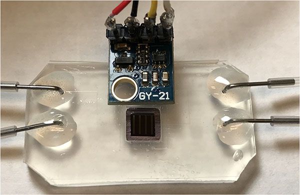 |
| The fluid-holding device designed by Holman and Zhao, which is held in front of an infrared beam to study the change in atomic bonds in sample of molecules. (Image: Hoi-Ying Holman, Berkeley Lab)
|
|
The results from this method, a type of operando spectroscopy methodology that uses Fourier transform infrared spectromicroscopy, indicate that the hydrogen bonds in the fibrils are indeed acting as roadblocks for the enzymes.
|
|
“Until now, the enzymatic hydrolysis of cellulose was a challenging biochemical process to study, in part due to limitations in researchers’ ability to carefully control, and observe, the sample environment during cellulose depolymerization,” said Hoi-Ying Holman, who is a senior scientist in the Lab’s Biosciences Area and BSISB director.
|
|
Holman, who co-led the project with Jeoh, designed this experimental system with then BSISB postdoc Wujun Zhao. She said, “This technology is the result of many years of research and development work carried out by BSISB staff to create systems for studying reactions using infrared spectromicroscopy.”
|
|
According to Holman, the team’s new approach overcomes past limitations thanks to the exceptional brightness of the ALS’ infrared beam, which allows for precise, real-time snapshots of the sample. Secondly, the fluid-holding device is partially open rather than sealed shut, enabling on-demand access to the sample while maintaining the necessary conditions for the enzymatic reaction and data collection.
|
|
“These results serve as a key proof-of-concept for the technique, with the potential to enable a new generation of scientific discoveries – especially for researchers hoping to study, and engage with, physical and chemical properties of biological systems as they occur in their native environments and in real time,” said Holman.
|
|
Scientists at LLNL plan to use the approach to study biomolecules in soil and plant and animal tissues, particularly those with biosecurity applications. Meanwhile, Jeoh and her team at UC Davis plan to explore techniques that will help enzymes break through the hydrogen bonds more quickly, to improve efficiency and lower costs of sustainable biomanufacturing.
|

