| Posted: Jul 27, 2009 |
Cellular toxicity of titanium dioxide nanotubes and nanowires |
| (Nanowerk Spotlight) One of the complications of nanotoxicology is that the toxicity of a specific nanomaterial cannot be predicted from the toxicity of the same material in a different form. For instance, while the toxicity of inert systems such as iron oxides, gold, or silver has been investigated for nearly isotropic particles (i.e., with a low aspect ratio), the toxicity of these materials in nanofilament form cannot be predicted from their known toxicity as nanoparticles. Fully understanding the toxic mechanisms of nanoscale materials is an essential prerequisite in being able to design harmless nanomaterials whose interactions with biological cells is non-lethal. |
| Currently, a lot of nanotoxicological research effort is focused on carbon nanotubes, but nanofilaments are not exclusively based on carbon materials and can be produced from many inorganic materials in the form of nanotubes and nanowires. Applying the 'precautionary principle' to nanotechnology would require much more extensive nanotoxicological research on all types of nanomaterials; and there seems to be a particular lack of findings concerning non-carbon nanofilaments. Researchers in Switzerland have now taken a closer look at the fate of titanium dioxide (TiO2) based nanofilaments in the body. Their results are cause for concern. |
| "TiO2 nanoparticles are widely used as UV blockers in sunscreens" says Arnaud Magrez. "Their cytotoxicity has been tested before and they were found to be rather non-toxic. Our new study shows that TiO2 based nanofilaments, however, can be quite toxic. The geometry of nanoparticles appears to play a crucial role in cytotoxicity. Furthermore, the toxicity can be enhanced by the presence of defects on the nanofilament surface, resulting from chemical treatment." |
| These new findings clearly demonstrated that the presence of TiO2 nanofilaments (synthesized by hydrothermal treatment from anatase and highly concentrated NaOH solution) had a strong dose-dependent effect on cell proliferation and cell death. Nanofilament internalization and alterations in cell morphology were observed. Acid treatment performed to substitute Na+ with H+ in the nanofilaments strongly enhanced the cytotoxic action. |
| Magrez, a researcher at the NN Research Group (Laboratoire de nanostructures et nouveaux matériaux électroniques) at the Ecole Polytechnique Fédérale de Lausanne (EPFL) in Switzerland, is first author of a recent paper in ACS Nano ("Cellular Toxicity of TiO2-Based Nanofilaments"). In this work, Magrez and colleagues from his group as well as the University of Fribourg, studied the cellular toxicity of TiO2-based nanofilaments in relation to their morphology and surface chemistry. |
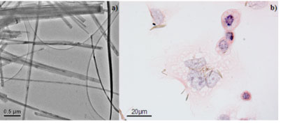 |
| Transmission Electron micrograph of TiO2-based nanowires. (a) Irreversible injuries and several morphological alterations of H596 epithelial cells can be observed after 2 days exposure with TiO2-based nanofibers (concentration is 2µg per ml). TiO2-based Nanofibers appear as golden wires (b). (Image: Arnaud Magrez, EPFL) |
| Apart from wide-spread use of TiO2 nanoparticles, large-scale arrays of TiO2-based nanofilaments (including nanotubes and nanowires) are already being used in photovoltaic cells and in photoelectrolyzer for the production of hydrogen by water splitting. |
| The surface cells of the airways, including the epithelial cells of the lungs, are the first cell type to encounter TiO2-based nanofilaments released into the environment. Therefore the Swiss researchers decided to investigate the acute cytotoxicity of different TiO2-based nanofilaments on lung cells in vitro. In their experiments they evaluated the cytotoxic effect of the TiO2-based nanofilaments by the widely established MTT assay performed with H596 human lung tumor cells. |
| Because the MTT assay measures the combined effects of cell proliferation and metabolic activity of cells and was reported to be prone to artifacts under certain experimental conditions, the EPFL team also validated their results by directly counting the number of surviving cells from microphotographs. Magrez says that, in comparison to untreated cells, both the MTT signals and number of cells were decreased in all nanomaterial-treated samples and the two methods – MTT assay and cell counting – yielded essentially identical results. |
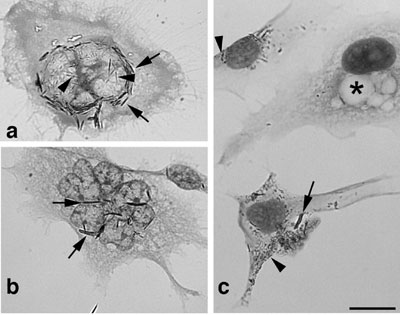 |
| Morphology of H596 lung carcinoma cells exposed to TiO2-based nanofilaments. Cells were treated for 4 days with 2 µg/mL TiO2 nanotubes, fixed and HE-stained. (a) Nanofilaments inside the cells have needle-like structures often concentrated around the enlarged nucleus (arrows), and even thinner nanofilaments are found within the nucleus (arrowheads). (b) In some giant cells, the nuclei are strongly lobulated or possibly fragmented, and the nanofilaments are localized between the lobules (arrows). In (a) and (b), a weaker staining of the nuclei was chosen to better visualize the intranuclear nanotubes. (c) Besides larger fibers (arrow), nanofilaments are also present in the form of small dark particles (arrowheads). Scale bar: 20 µm. (Reprinted with permission from American Chemical Society) |
| The researchers found that geometry of the nanomaterials appears to play a role, surface chemistry was the most important aspect in determining the survival of exposed cells. |
| "The importance of surface chemistry had already been observed in a previous report on carbon-based nanomaterials," says Magrez. "Even though multiwalled carbon nanotubes have comparable diameter and length as TiO2 nanofilaments, their toxicity is markedly different. Thus, the chemical composition of nanomaterials also appears to have an effect on cell survival. Whether the toxicity determined in this acute model will also translate to models of chronic toxicity or even tumor development (lung carcinoma or mesotheliomas) needs to be addressed in future studies." |
 By
Michael
Berger
– Michael is author of four books by the Royal Society of Chemistry:
Nano-Society: Pushing the Boundaries of Technology (2009),
Nanotechnology: The Future is Tiny (2016),
Nanoengineering: The Skills and Tools Making Technology Invisible (2019), and
Waste not! How Nanotechnologies Can Increase Efficiencies Throughout Society (2025)
Copyright ©
Nanowerk LLC
By
Michael
Berger
– Michael is author of four books by the Royal Society of Chemistry:
Nano-Society: Pushing the Boundaries of Technology (2009),
Nanotechnology: The Future is Tiny (2016),
Nanoengineering: The Skills and Tools Making Technology Invisible (2019), and
Waste not! How Nanotechnologies Can Increase Efficiencies Throughout Society (2025)
Copyright ©
Nanowerk LLC
|

Become a Spotlight guest author! Join our large and growing group of guest contributors. Have you just published a scientific paper or have other exciting developments to share with the nanotechnology community? Here is how to publish on nanowerk.com. |
