| Posted: Apr 26, 2007 |
Watching carbon nanotubes grow |
| (Nanowerk Spotlight) Synthesis of carbon nanotubes (CNTs) is a rapidly advancing field, but there is a lot that researchers don't know about how nanotubes form and grow. Synthesis, while rapidly developing, is currently the weakest link for most nanotube applications, with high yield and high precision diameter and chirality control being important goals. Historically, in situ characterization tools have accelerated progress in synthesis for many advanced materials, and there is widespread recognition that in situ tools have the potential to improve CNT synthesis as well. Ideally one would like to detect individual nanotubes and ensembles as they grow and measure their physical properties while imposing minimal constraints on the synthesis method. In other words, with a good understanding of the synthesis process we would be better able to control the product. It is feasible that by actually observing nanotubes as they grow one will gain a better understanding of the growth process and also better characterize the grown product. Greater control over the physical characteristics of the nanotube product is essential to enable many applications, as well as many fundamental studies. Although chemical vapor deposition (CVD) is now a very standard method to synthesize CNTs, there aren't really standard in situ tools to characterize nanotubes during growth. Researchers in Canada have now shown how global Raman imaging (GRI) can be used to characterize the CVD growth of CNTs in situ and in real time. |
| "We have been developing methods to optically image individual carbon nanotubes" Dr. Paul Finnie tells Nanowerk. "In our recent work we show that we can capture spatially resolved optical images of many nanotubes or even one single-walled carbon nanotube inside the reactor in which they are synthesized, despite their nano scale and despite adverse conditions such as high temperatures." |
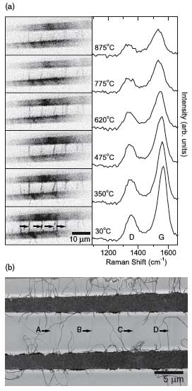 |
| Raman imaging at various temperatures. (a) The left column shows Raman images taken at different temperatures and the right column shows the corresponding Raman spectra showing G band and D band evolution with temperature. (b) A room temperature SEM image of the same area taken at 1 kV. The contrast on the image is inverted for better viewing. The black arrows indicate suspended nanotubes which are also visible in the Raman image. (Reprinted with permission from IOP Publishing) |
| Finnie is a researcher in the Quantum Physics Group at the Institute for Microstructural Sciences, National Research Council of Canada, in Ottawa. Together with his colleagues he is working on ways to better control synthesis of CNTs. |
| "It is my view that one of the best ways to gain control over synthesis is to watch it happen" says Finnie. "We are developing tools to do this.In the short term we can learn a lot about the dynamics of how nanotubes nucleate and grow. In the longer term we could imagine this as a standard way to monitor CVD growth of nanotubes." |
| Finnie points out that there are a number of in situ characterization methods for nanotube synthesis under development worldwide, each with different strengths and weaknesses. For instance,it is well established that electron beams and electric fields are at least invasive for nanotubes, and at worst highly destructive. Furthermore, electron microscopes are prohibitively expensive and difficult to adapt to standard CVD reactors. In another approach that is less costly and more closely adapted to standard CVD, electron field emission has been imaged during the cold walled synthesis of CNTs. However, much of the information about the nanotube structure is indirect and again, fields and currents are invasive. |
| There are also several non-imaging in situ characterization methods, and while some may potentially be pushed to the single nanotube level, to date they have been applied only to ensembles. |
| Thie method used by Finnie and his collaborators is an all-optical method that is simple, versatile, nondestructive, non-invasive and economical. Called Global Raman Imaging (GRI), this method obtains an image formed by spatially resolving the scattered light from a Raman band of interest. It is complementary to conventional Raman spectroscopy, and enables observations down to the single nanotube level to be made in situ and in real time. Finnie's team were the first to apply GRI to nanotubes, especially SWNT, and the first to use it with nanotube CVD. |
| Finnie cautions that the method should not be confused with more conventional confocal Raman microscopy, in which a series of Raman spectra are taken in a raster pattern to build up a ?hyperspectral? image. "GRI, as used here, does not supply spatially resolved spectroscopic data, which is a limitation of the technique compared to conventional Raman raster scanning" he says. "Instead of detailed spectral information, here we use GRI to provide rapid spatial resolution of a single spectral feature of interest." |
| In GRI, the sample is illuminated by a defocused spot, and imaged directly, with the spectral band of interest selected by a filter. This enables an instantaneous snapshot to be taken of the spatial distribution of any given Raman signal. |
| "Importantly, because the spot size is relatively large, nanotubes need never be exposed to a strong optical field, which we have shown can manipulate suspended CNTs" says Finnie. |
| Optical imaging of single-walled CNTs has only developed in the past couple of years. Researchers have just begun to explore this area and it still is a wide open territory for research. Finnie says the field of nanotube optics is "really blossoming right now." He is co-organizing a workshop on this topic this spring in Ottawa (WONTON'07 - 2nd workshop on Nanotube Optics & Nanospectroscopy). |
| A lot of nanotube research is sample driven says Finnie. "Nanotube sample quality is improving very quickly worldwide by a lot of measures. Research is expanding into many directions. Right now there is a lot of basic research, and fantastic progress, but in my view the field today is weaker in applications then it should be. In addition to the beautiful basic research I'd like to see several very practical "killer apps" emerge, and I think they will pretty soon." |
| The research, titled "Real-time in situ Raman imaging of carbon nanotube growth", has been published in the March 23, 2007 online edition of Nanotechnology. It was funded by a JST grant as part of the "Nanofactory" research program organized by Prof. Y. Homma. |
 By
Michael
Berger
– Michael is author of four books by the Royal Society of Chemistry:
Nano-Society: Pushing the Boundaries of Technology (2009),
Nanotechnology: The Future is Tiny (2016),
Nanoengineering: The Skills and Tools Making Technology Invisible (2019), and
Waste not! How Nanotechnologies Can Increase Efficiencies Throughout Society (2025)
Copyright ©
Nanowerk LLC
By
Michael
Berger
– Michael is author of four books by the Royal Society of Chemistry:
Nano-Society: Pushing the Boundaries of Technology (2009),
Nanotechnology: The Future is Tiny (2016),
Nanoengineering: The Skills and Tools Making Technology Invisible (2019), and
Waste not! How Nanotechnologies Can Increase Efficiencies Throughout Society (2025)
Copyright ©
Nanowerk LLC
|

Become a Spotlight guest author! Join our large and growing group of guest contributors. Have you just published a scientific paper or have other exciting developments to share with the nanotechnology community? Here is how to publish on nanowerk.com.
