| Posted: Jul 30, 2007 |
Nanodiamonds as alternative to toxic quantum dots |
| (Nanowerk Spotlight) Recent developments in spectroscopic techniques allow highly sensitive image detection both in vitro and in vivo on the individual cell level. These methods depend on nanometer-size particles as detection probes. One class of such particles, so-called nanocrystals or quantum dots (qdots), is very popular for constructing detection probes for biolabeling. Scientists have discovered that these nanocrystals can enable researchers to study cell processes at the level of a single molecule and may significantly improve the diagnosis and treatment of diseases such as cancers. Qdots are either used as active sensor elements in high-resolution cellular imaging, where the fluorescence properties of the qdots are changed upon reaction with the analyte, or in passive label probes where selective receptor molecules such as antibodies have been conjugated to the surface of the dots. Qdots could revolutionize medicine. Unfortunately, most of them are toxic. Ironically, the existence of heavy metals in qdots such as cadmium, a well-established human toxicant and carcinogen, poses potential dangers especially for future medical application, where qdots are deliberately injected into the body. As the use of nanomaterials for biomedical applications is increasing, environmental pollution and toxicity have to be addressed, and the development of a non-toxic and biocompatible nanomaterial is becoming an important issue. Researchers are now proposing the use of nanoscale diamond particles as a non-toxic alternative to heavy metal qdots. |
| "We have shown that surface-modified carboxylated nanometer-sized diamond (cND) can be easily detected by bio-AFM, confocal microscopy and flow cytometry" Dr. Jui-I Chao explains to Nanowerk. "Importantly, the existence of cNDs inside a cell does not induce cell death. The biocompatible and detectable properties of cNDs are suitable for further biomedical applications including bio-labeling, imaging and drug delivery." |
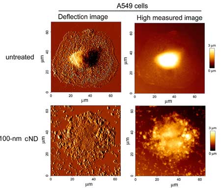 |
Observation of 100 nm cND interacted with cell by Bio-AFM. A549 cells were treated with or without 50 µg ml-1 100 nm cNDs for 4 hours. After cell fixation, the cells were observed by Bio-AFM. The upper pictures indicated the cND-untreated cell. The lower pictures showed that the cell was treated with 100 nm cND. (Reprinted with permission from IOP Publishing) |
| Chao, a Professor of Pharmacology and Toxicology at Tzu Chi University in Taiwan, together with a group of researchers from other Taiwanese universities, has recently published a paper in which they demonstrated the advantages of nanodiamond’s unique properties with its biocompatibility, nontoxicity, and natural fluorescence without complicated pretreatments ("Biocompatible and detectable carboxylated nanodiamond on human cell"). |
| It has been previously reported ("High-Affinity Capture of Proteins by Diamond Nanoparticles for Mass Spectrometric Analysis") that the surface of nanodiamonds provides a unique platform for bio-conjugation after chemical modifications. For instance, nanodiamonds whose surface was modified by carboxylation exhibited high affinity for proteins. |
| In the course of the study, various sizes of cNDs were evaluated with regard to their cytotoxicity in human lung cells. "We accumulated 5 nm and 100 nm size cND particles on A549 lung epithelial cells and HFL-1 lung fibroblast" says Chao. "We found that these cND particles did not induce cytotoxicity and apoptosis. Interestingly, both 5 nm and 100 nm cNDs display natural green fluorescence upon laser excitation in cells that can be detected by scanning confocal microscopy and flow cytometry. Recently, high energy treated nanodiamond was found to emit bright fluorescence and no photobleaching was detected." |
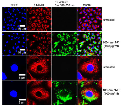 |
Detection of cNDs in A549 cells by a laser scanning confocal microscope. A549 cells were treated with or without 100 µg ml-1 100 nm cNDs for 4 hours and then recovery for 24 hours. After cell fixation, the cells were incubated with the Cy3-labelled antiβ-tubulin. The green light signal from the cNDs was excited with wavelength 488 nm and the emission was collected in the range of 510–530 nm. The β-tubulin protein displayed red fluorescence. The nuclei were stained with Hoechst 33258, which displayed blue fluorescence. (Reprinted with permission from IOP Publishing) |
| Chao concludes that the biocompatible and detectable properties of cND nanoparticles should therefore be considered as a novel and relatively safe nanomaterial for further biomedical applications. |
| In contrast to the cNDs, the researchers found in their experiments that carbon nanotubes induced cytotoxicity in human lung cells. |
| While this paper investigates the toxicity and biocompatibility in a lung cellular model, the potential human health effects from exposure to nanodiamonds need to be determined by further in vivo tests in animals. Especially the mechanisms of transportation, biotransformation, and metabolism of nanodiamond particles in animals need further investigation. |
| If it indeed turns out that nanodiamonds are a benign material, the development of nanodiamonds conjugated with biological molecules and chemical drugs for biomedical applications appears to have a bright future. Nanodiamonds could potentially be used as novel probes for both diagnostic and therapeutic applications. |
| "The modified surface of carbon nanoparticles may be conjugated with biomolecules such as specific proteins or peptides for the detection of specific cells or tissues (e.g. cancer cells)" says Chao. "Furthermore, nanodiamond conjugated with specific drug molecules might be used for the therapy of patients." |
 By
Michael
Berger
– Michael is author of four books by the Royal Society of Chemistry:
Nano-Society: Pushing the Boundaries of Technology (2009),
Nanotechnology: The Future is Tiny (2016),
Nanoengineering: The Skills and Tools Making Technology Invisible (2019), and
Waste not! How Nanotechnologies Can Increase Efficiencies Throughout Society (2025)
Copyright ©
Nanowerk LLC
By
Michael
Berger
– Michael is author of four books by the Royal Society of Chemistry:
Nano-Society: Pushing the Boundaries of Technology (2009),
Nanotechnology: The Future is Tiny (2016),
Nanoengineering: The Skills and Tools Making Technology Invisible (2019), and
Waste not! How Nanotechnologies Can Increase Efficiencies Throughout Society (2025)
Copyright ©
Nanowerk LLC
|

Become a Spotlight guest author! Join our large and growing group of guest contributors. Have you just published a scientific paper or have other exciting developments to share with the nanotechnology community? Here is how to publish on nanowerk.com.
