| Posted: Nov 08, 2011 |
When your food makes you glow - a novel optical bioimaging technique |
| (Nanowerk Spotlight) Fruit flies (Drosophila melanogaster) are the workhorses in countless biomedical research laboratories around the world. What makes these insects so valuable as model organisms for scientists is that they possess strikingly similar versions of the genes that promote normal human development and, when altered, contribute to disease. |
| The bioimaging of live specimens, ideally through all the stages of the fruit fly life cycle, is a tricky and often complicated undertaking. Magnetic resonance imaging, for instance, can be time consuming, costly, and often burdened with operational difficulties – the handling of flies inside the NMR tube has been described as a very tricky job. Another option is optical imaging using fluorescent nanomaterials as contrast agents. These need to be introduced by intravenous/intramuscular injection or by gene translation, both of which can be difficult and time consuming. Also, genetically modified fluorescence proteins tend to be not very stable. |
| Another aspect here is the varying degree of toxicity of these agents – semiconductor-based quantum dots; silicon nanoparticles; or surface-passivated carbon dots. |
| Researchers in India have now developed a relatively simple way to introduce fluorescent nanomaterials into fruit flies: They prepared carbon nanoparticles from wood waste and added them to the flies' food supply. As they reported in the October 20, 2011 online edition of Small ("Carbon Nano-onions for Imaging the Life Cycle of Drosophila Melanogaster"), just 4 ppm of these nanoparticles allowed the optical fluorescence microscopy imaging of all the stages of the fruit fly life cycle. The fluorescent fruit flies showed no toxic effects – upon withdrawal of the nanoparticles from their food, they excreted the fluorescing material and continued to proliferate to the next generation, demonstrating a return to their normal lives. |
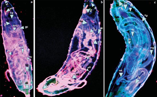 |
| Images (merges of 385, 488, and 561 nm fi lters) of Drosophila melanogaster at the larval stages showing organ details: a) fi rst instar larva, b) second instar larva, and c) third instar larva. Organ labels: at = atrium; px = pharynx; sd = salivary duct; sg = salivary gland; ep = esophagus; pvc = proventiculus; gc = gastric ceaca; mg = midgut; mi = mid intestine; utr = ureter; mt = malpighian tubule; hg = hind gut; as = anus. (Reprinted with permission from Wiley-VCH Verlag) |
| "Our results demonstrate that simple, noninvasive oral ingestion of fluorescent carbon nano-onions enables the imaging of the full life cycle, including larval organs, of Drosophila melanogaster in all of its developmental stages," Sabyasachi Sarkar, Professor of Chemistry at the Indian Institute of Technology in Kanpur, tells Nanowerk. "The intensity of this fluorescent material is increased in vivo." |
| These findings by Sarkar and his team finding open up the prospect of optical imaging of living species in a relatively simple, non-toxic and cost effective way. In addition, colorful imaging is possible to render better contrast without the use of any perishable external organic coloring molecules or fluorescence proteins. |
| The researchers avoided exotic methods like laser ablation to fabricate their nanocarbon and instead followed the primitive method of burning carbonaceous materials like wood wool as used long ago by Thomas Edison for the carbon filament for his light bulb. That way they synthesized fluorescent, water-soluble carbon nano-onions by carbonizing wood waste under pyrolysis. They followed this by surface oxidation to isolate their nanoparticles. These were then uniformly mixed with the food of Drosophila. |
| "The nano-onions are heavily derivatized upon oxidation, with carboxylic acid groups on the surface rendering them water-soluble" explains Sarkar. "These are selectively filtered through micropore filters to get the smallest possible sizes, which are microscopically analyzed before use." |
| He notes that the advantage of this multishell carbon nano-onions for derivatization (oxidation) serves several important points: "Firstly, these nano-onions are defective, so oxidation is readily made with the incorporation of high density carboxylic groups on the outer shells, transforming these hydrophobic carbon into a very hydrophilic material. Secondly, the surface passivation is made to exhibit visible luminescence. Thirdly, further passivation in vivo is possible when the surface carboxylate groups interact with several biomolecules available in the species. Therefore these carbon nano onions are cheaply made, reproducible, and enhance the fluorescence activity in vivo." |
| Toxicological investigations by the team showed no adverse effects on the flies. Once the carbon nanoparticles were removed from their food, the flies gradually lose fluorescence by excretion, demonstrating that carbon nano-onions are readily removed from the body. |
| Quantification of the association of carbon nano-onions with several common biomolecules to effect the fluorescent enhancement and hence the imaging of the life cycle of zebra fish is currently in progress. This technique may be used in the easy, noninvasive image-based studies of other animals, and could ultimately be extended to human beings. |
| Another important aspect of this work, as Sarkar points out, is that these carbon nano-onions can be used as drug carrier molecules, and the entire drug delivery process can be developed and visualized. |
 By
Michael
Berger
– Michael is author of four books by the Royal Society of Chemistry:
Nano-Society: Pushing the Boundaries of Technology (2009),
Nanotechnology: The Future is Tiny (2016),
Nanoengineering: The Skills and Tools Making Technology Invisible (2019), and
Waste not! How Nanotechnologies Can Increase Efficiencies Throughout Society (2025)
Copyright ©
Nanowerk LLC
By
Michael
Berger
– Michael is author of four books by the Royal Society of Chemistry:
Nano-Society: Pushing the Boundaries of Technology (2009),
Nanotechnology: The Future is Tiny (2016),
Nanoengineering: The Skills and Tools Making Technology Invisible (2019), and
Waste not! How Nanotechnologies Can Increase Efficiencies Throughout Society (2025)
Copyright ©
Nanowerk LLC
|

Become a Spotlight guest author! Join our large and growing group of guest contributors. Have you just published a scientific paper or have other exciting developments to share with the nanotechnology community? Here is how to publish on nanowerk.com. |
