| Posted: Feb 28, 2006 |
Nanoparticle-stabilized liposomes |
| (Nanowerk News) Liposomes, man-made cells used as drug delivery vehicles, would be more useful if only they could be stabilized against fusion with one another. Researchers at the University of Illinois told Nanowerk that they succeeded in doing exactly that - they stabilized phospholipid liposomes with charged nanoparticles, thereby opening up interesting functional perspectives. |
| Liposomes, which were first proposed and tested as a drug delivery system some 30 years ago, are artificially constructed spherical cells of phospholipid bilayers. Liposomes are useful for two reasons: |
| Firstly, because they are hollow, they can be used as delivery vehicles for a wide variety of drugs, vaccines, enzymes, nonenzyme proteins, and genetic material. These nanocontainers within liposomes have volumes from zeptoliters to femtoliters. To release the "payload" one can either change the temperature to a certain threshold level or use pulses of a strong electric field to break the cell membrane apart. The cell then spills its contents into the bloodstream or into tissues to which it has migrated by diffusion through the walls of capillaries. |
| Secondly, liposomes are tremendously biofunctionalizable; antibodies, protein receptors, and other biosensor molecules can attach to their outer surface. |
| A problem with using liposomes as delivery vehicle is their stability: When liposomes encounter one another in suspension, they are prone to adhere and fuse to form a larger one. Moreover, when liposomes open during the fusion process the result could be inclusion leakage, unexpected mixing between chemicals that are nominally separated into different liposome capsules, and inefficient reactions. |
| Professor Steve Granick of the University of Illinois, and one of his students, Liangfang Zhang, came up with a simple strategy of mixing phospholipid liposomes with charged nanoparticles (carboxyl-modified polystyrene) and using sonication to mix them at low volume fraction to produce particle-stabilized liposomes that repel one another and do not fuse. |
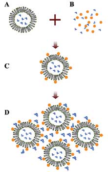 |
| Schematic illustration of the experimental strategy to produce particle-stabilized liposomes. (A) DLPC liposomes with a diameter of 200 nm were made using the extrusion method. (B) Carboxyl-modified polystyrene nanoparticles with a diameter of 20 nm were prepared. (C) Nanoparticle-stabilized liposomes were formed by mixing A and B at the molar concentration ratio of 1:100, with 10 min of sonication. (D) To condense the dilute liposome suspension C, nitrogen gas was blown gently over the suspension until the desired volume fraction was attained. (Source: University of Illinois, Professor Granick) |
| An ideal stabilization method would leave untouched a substantial fraction of the liposome surface, leaving a substantial portion of phospholipid surface area free for functionality. Granick and Zhang showed that their nanoparticle stabilization process meets this need with an estimated free membrane outer surface, not covered with nanoparticles, of ∼75%. |
| As Granick told Nanowerk "these nanoparticle-stabilized liposomes may be interesting from several functional perspectives." |
| "First, they comprise potentially a novel kind of colloidal-sized sensor. Antigens and membrane proteins can be embedded within the large free area contained within them; yet because they resist fusion with one another, they can be concentrated to exceptionally high volume fractions and consequently to exceptionally high functionality when the ratio of surface-to-volume is large." |
| "Second, we imagine that chemical reactions can be performed within these objects at the individual liposome level. Because each liposome is impermeable, this signifies that elementary reactions, each of them performed at dilute concentration, can be studied in parallel among many such objects." |
| "And third, these objects afford a new type of colloidal particle: hollow, deformable, and biofunctionalizable. Their rheology, diffusion, and potential crystallization may contribute to the study of structure and dynamics of colloidal-sized objects, at very high volume fractions where particle-particle interactions dominate, in the case where the particle contour is more "raspberry" than spherical." | The research paper, titled "How to Stabilize Phospholipid Liposomes (Using Nanoparticles)" was published in the Feb. 22, 2006 online edition of Nano Letters. |
 By
Michael
Berger
– Michael is author of four books by the Royal Society of Chemistry:
Nano-Society: Pushing the Boundaries of Technology (2009),
Nanotechnology: The Future is Tiny (2016),
Nanoengineering: The Skills and Tools Making Technology Invisible (2019), and
Waste not! How Nanotechnologies Can Increase Efficiencies Throughout Society (2025)
Copyright ©
Nanowerk LLC
By
Michael
Berger
– Michael is author of four books by the Royal Society of Chemistry:
Nano-Society: Pushing the Boundaries of Technology (2009),
Nanotechnology: The Future is Tiny (2016),
Nanoengineering: The Skills and Tools Making Technology Invisible (2019), and
Waste not! How Nanotechnologies Can Increase Efficiencies Throughout Society (2025)
Copyright ©
Nanowerk LLC
|

Become a Spotlight guest author! Join our large and growing group of guest contributors. Have you just published a scientific paper or have other exciting developments to share with the nanotechnology community? Here is how to publish on nanowerk.com.
