| Posted: May 08, 2006 |
Towards single-particle laboratories |
| (Nanowerk Spotlight) Researchers at Cornell University have developed a novel quantitative nanoparticle-based sensor of chemical concentrations based on organic dye molecules covalently integrated into the matrix of silica nanoparticles. This is the first work that implements an optimized core-shell architecture for such sensor particles. |
| The Cornell researchers' findings are the cover article of the June 2006 issue of Small: "Core/Shell Fluorescent Silica Nanoparticles for Chemical Sensing: Towards Single-Particle Laboratories". |
| These particles build on previous research in the Wiesner Research Group in Cornell's Department of Materials Science and Engineering. This work showed that encapsulation of dyes within a core-shell particle architecture can significantly enhance the brightness and stability of the dye molecules within. |
| "To develop our core-shell sensor particles, we have encapsulated a reference dye species within 50nm sol-gel silica core particles, which are then coated in a 10 nm thick surface shell which contains an environmentally-sensitive sensor dye" Andrew Burns, the paper's first author explained to Nanowerk. |
| "By comparing the emission of both sensor and reference dyes, we are able to determine the concentration of an analyte, independent of the sensor concentration" Burns says. "Thus, we have developed an optimized sensor geometry, placing the reference dye in the core, away from perturbations, while exposing the sensor dye to the maximum possible surface area for interaction with analytes." |
| The silica matrix also acts to protect both the sensor and reference dyes from interactions with solvent or other molecules which could alter the dyes' fluorescence, while allowing small molecule analytes to diffuse in and out easily. The concept is initially demonstrated for sensing pH, and has been performed both in solution as well as within cells. |
| The novelty of this work is the core-shell geometry. "By placing the sensor and reference dyes in separate shells, we (a) preclude energy transfer between them, as well as (b) protecting the reference dyes and (c) placing the sensor dyes where they are most exposed to the environment" Burns explains. |
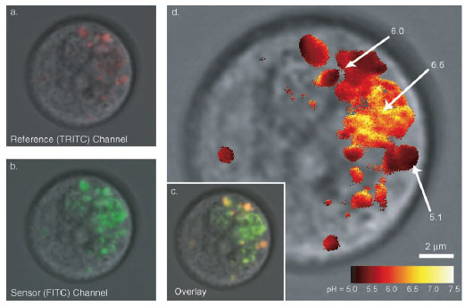 |
| Confocal fluorescence microscopy images (overlaid on bright field) of pH sensors in RBL mast cells showing a) reference dye channel, b) sensor dye channel, c) overlaid images, and d) false-color ratiometric imaging of pH in various intracellular compartments. (Reprinted with permission from Wiley) |
| The Cornell group's work addresses the specific problem of quantitative sensing of an analyte on a very short length scale and within a complex environment (e.g. within a living cell). Unlike macroscopic environments, where tests such as litmus paper or electrochemical cells can be used to detect changes in pH, one must create a very small sensor to be able to analyze ion concentration without perturbing the biological system under investigation. |
| "Nanoparticles, such as the core-shell fluorescent silica particles we have developed, are ideal probes for intracellular studies because of their small size and biocompatible silica matrix" says Burns. "Furthermore, the small particle size yields very high spatial and temporal resolution to monitor local conditions at the nanoscale." |
| This research could potentially be applied to a variety of questions in biology and pharmacology. For example, "cocktails" of particles with various sensing capabilities (intracellular analytes such as Ca+2, reactive oxygen, etc) could be applied simultaneously in vitro to determine the effects of novel therapeutics on intracellular ion concentrations and to generate high-content screens for metabolic and cellular health status, providing pharmacologists with a wealth of information to allow them to create more effective treatments. |
| Looking forward, Burns says: "I believe that the future of the field of nanobiotechnology as a whole and the field of core-shell silica nanoparticles in particular lies in the development of multifunctional materials which bring together many capabilities which may have been previously realized separately, onto one nano-sized vehicle. For our work, silica is ideally suited to this development, as it is a very versatile host material which has already shown the ability to integrate fluorescence (for imaging and sensing), ordered mesoporosity (for high surface area and/or controlled drug release), magnetism (for MRI imaging and separation/mobility), biomolecules (for targeting), and many other capabilities." |
| By bringing together multiple functionalities in separate shells of a single particle, it may be possible one day to develop novel "single-particle laboratories" which could facilitate research in a variety of fields from fundamental biology to pharmacology and beyond. |
 By
Michael
Berger
– Michael is author of four books by the Royal Society of Chemistry:
Nano-Society: Pushing the Boundaries of Technology (2009),
Nanotechnology: The Future is Tiny (2016),
Nanoengineering: The Skills and Tools Making Technology Invisible (2019), and
Waste not! How Nanotechnologies Can Increase Efficiencies Throughout Society (2025)
Copyright ©
Nanowerk LLC
By
Michael
Berger
– Michael is author of four books by the Royal Society of Chemistry:
Nano-Society: Pushing the Boundaries of Technology (2009),
Nanotechnology: The Future is Tiny (2016),
Nanoengineering: The Skills and Tools Making Technology Invisible (2019), and
Waste not! How Nanotechnologies Can Increase Efficiencies Throughout Society (2025)
Copyright ©
Nanowerk LLC
|

Become a Spotlight guest author! Join our large and growing group of guest contributors. Have you just published a scientific paper or have other exciting developments to share with the nanotechnology community? Here is how to publish on nanowerk.com.
