| Posted: Aug 28, 2006 |
Infrared imaging of sub-10 nm particles |
| (Nanowerk Spotlight) Objects a thousand times smaller than the wavelength of infrared light (10 micrometers) are undetectable by standard far-field optical infrared microscopy since the weak nanoparticle signals would be buried far below the background level. To overcome this drawback and to achieve nanoscale spatial resolution researchers in Germany illuminate the nanoparticles by a highly intensive nanoscale infrared light spot. It is generated in the nano-gap between a laser-illuminated scanning metal tip and the substrate supporting the particles. The simple but very efficient trick finally allowing detection of sub-10 nm particles is the use of highly reflecting substrates instead of glass slides typically used as a sample carrier in optical microscopy. |
| There is still a strong need in material-specific methods for chemical identification of nanocomposite materials. While the classical optical and infrared microscopies are highly sensitive to the chemical composition of materials, the diffraction limit prevents imaging of nanoscopic details, like the electron microscope does. In addition to the high chemical sensitivity, light microscopy offers several further advantages which are essential for applications in biology and biochemistry: it is nondestructive, radiation damage is negligible and objects can be imaged in liquids. |
| Researchers at the Max Planck Institute (MPI) of Biochemistry near Munich, Germany, are developing a new optical microscopy that allows a significantly better resolution than conventional optical microscopy. The microscope is based on an atomic force microscope (AFM) where the scanning tip is used for both mechanical and near-field optical probing. They call this scattering-type scanning near-field optical microscopy, s-SNOM. The laser-illuminated tip concentrates the light at the very tip apex thereby generating an optical nanofocus scanning the sample. It was already shown that a resolution at the nanometer-scale can be achieved, up to 1000 times better than in conventional infrared microscopy. |
| New experiments by the MPI researchers enable for the first time infrared microscopy of single gold nanoparticles with diameters as small as 8 nm at wavelengths of about 10 µm. |
| "We showed that the near-field optical particle contrast can be strongly enhanced by employing highly reflecting substrates such as gold and silicon" Rainer Hillenbrand, head of the Nano-Photonics Group at the institute explained to Nanowerk. The researchers found that particularly strong particle contrasts are achieved by resonant near-field coupling provided by phonon-polariton excitation in a silicon carbide substrate. |
| In a recent paper ("Infrared Imaging of Single Nanoparticles via Strong Field Enhancement in a Scanning Nanogap"), Hillenbrand and his colleagues describe how they apply near-field optical tip-substrate coupling to generate both strongly enhanced and confined optical fields for high-resolution near-field optical microscopy of nanoparticles. |
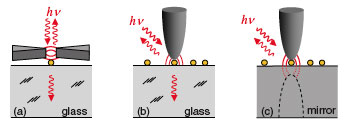 |
| Optical detection of nano-objects (yellow) employing the enhanced near fields (a) in the feed gap between two antenna arms, (b) at the apex of a metal probe tip, and (c) in the gap formed between a probe tip and a highly reflecting substrate. (reprinted with permission from the American Physical Society) |
| Usually, the objects to be imaged by a near-field probe are adsorbed on a low-dielectric substrate (e.g., glass) and the near-field coupling between tip and substrate is weak. |
| "By employing highly reflecting substrates we demonstrate that the infrared nanofocus illuminating the particles can be intensified" PhD-student Antonija Cvitkovic, first author of the work points out. "This effect is explained by the strong near-field coupling between tip apex and substrate, yielding highly concentrated optical fields for probing the particles"" |
| "A direct application of our method could be high-resolution imaging of gold-biolabels" says Hillenbrand. "Combined with spectroscopic mapping, our method opens the door to other exciting applications: noninvasive, label-free chemical identification of individual nanocrystals or biomolecules via their infrared fingerprint spectra". |
| Another exciting application, infrared near-field spectroscopy of organic and biological nanoparticles, was described in a recent article titled "Infrared Spectroscopic Mapping of Single Nanoparticles and Viruses at Nanoscale Resolution", published in the June 10, 2006 issue of Nano Letters. |
| Hillenbrand expects further improvement of the probe sensitivity by employing resonant and sharper probe tips, which could open the door to infrared spectroscopy of even single molecules at ultrahigh spatial resolution. Finally, by exploiting plasmon-polariton excitation in metal substrates the demonstrated effect could also push the limits in tip-enhanced spectroscopy at visible frequencies. |
 By
Michael
Berger
– Michael is author of four books by the Royal Society of Chemistry:
Nano-Society: Pushing the Boundaries of Technology (2009),
Nanotechnology: The Future is Tiny (2016),
Nanoengineering: The Skills and Tools Making Technology Invisible (2019), and
Waste not! How Nanotechnologies Can Increase Efficiencies Throughout Society (2025)
Copyright ©
Nanowerk LLC
By
Michael
Berger
– Michael is author of four books by the Royal Society of Chemistry:
Nano-Society: Pushing the Boundaries of Technology (2009),
Nanotechnology: The Future is Tiny (2016),
Nanoengineering: The Skills and Tools Making Technology Invisible (2019), and
Waste not! How Nanotechnologies Can Increase Efficiencies Throughout Society (2025)
Copyright ©
Nanowerk LLC
|

Become a Spotlight guest author! Join our large and growing group of guest contributors. Have you just published a scientific paper or have other exciting developments to share with the nanotechnology community? Here is how to publish on nanowerk.com.
