| Posted: Jul 31, 2015 |
Electron microscopy leads to discovery of new structural features of human hair
|
|
(Nanowerk News) A new discovery about the structure of human hair is likely to change the way scientists and researchers, as well as the cosmetics industry, view and explore it in the future.
|
|
Human hair structure has been studied extensively for more than 70 years, but a complete picture of its local structure has proven elusive.
|
|
But now, by combining a submicron X-ray beam with cross-section geometry, a team of researchers in Brazil and New York has detected new structural features of human hair. During the American Crystallographic Association (ACA) 2015 Meeting, which will be held in Philadelphia from July 25-29, Vesna Stanic, a scientist working at the Brazilian Synchrotron Light Source, will present an abstract, “Local and average structure of human hair,” describing their discovery and methods.
|
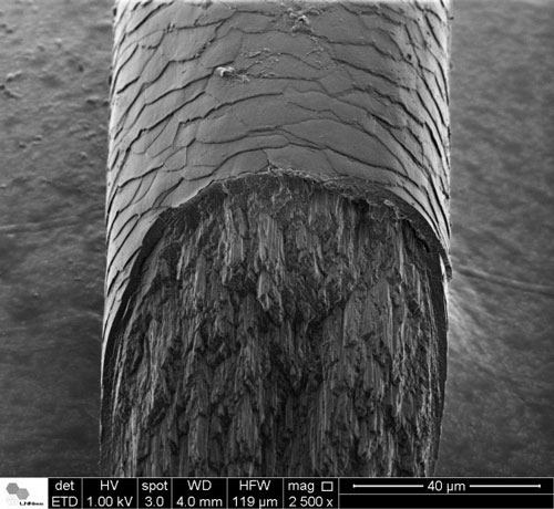 |
| Electron Microscopy micrograph of human hair cross-section. The top region shows the external part of the hair – cuticle region; The bottom region shows the internal macrofibrils – cortex region. (Image: Fabiano Emmanuel Montoro/LNNano, CNPEM)
|
|
If you’ve heard of “keratin,” you may already know that human hair consists primarily of keratin molecules arranged in a hierarchical sort of structure, in which the fundamental building block is known as an “intermediate filament.”
|
|
While studying materials used for hair treatments, Stanic wondered what effect these treatments were having on the diffraction pattern of the hair. X-ray diffraction patterns of a given material can reveal the local arrangement of molecular and atomic structure.
|
|
Although diffraction patterns have been examined and reported in several publications in the past, they involved bundles of hair fibers or microdiffraction on single hair fibers -- and, most significantly, the X-ray beam was always oriented perpendicular to the hair fiber axis.
|
|
So Stanic decided to take a closer look at the diffraction pattern of the hair by measuring it with an X-ray beam aimed parallel -- rather than perpendicular -- to the hair axis. “I wanted to use a submicron X-ray beam, so I asked colleague Kenneth Evans-Lutterodt to perform an experiment on the microdiffraction beamline at Brookhaven National Laboratory,” she added.
|
|
Using a submicron X-ray beam and transmission electron microscopy, they were able to spatially resolve the local structure of the three main regions of human hair: medulla, cortex and cuticle.
|
|
“We performed a full diffraction map from a 30-micron-thick cross section of hair, with an incident beam parallel to the hair axis and then compared it to the diffraction map with the beam perpendicular to the hair axis,” noted Stanic.
|
|
What they found
|
|
The researchers found an additional, previously unobserved structural region in the cortex near the cuticle boundary. “We also discovered that within the cuticle a key diffraction feature of the alpha keratin is absent -- indicating the presence of beta keratin instead of the alpha keratin phase,” said Stanic. Until now, it was believed that keratin in the whole hair had only an alpha conformation, she explained.
|
|
The work “provides irrefutable experimental evidence of the hair phase variation across the three main regions of hair and is an important step toward gaining a better understanding of hierarchical ordering of the intermediate filaments of keratin,” she said. “It also highlights the importance of using a submicron X-ray beam to unravel the structures of poorly ordered, multiphase systems such as hair.”
|
|
The team believes the cosmetics industry will benefit from their findings. “If the goal of the cosmetics industry is to make new and better products for hair, then it’s absolutely critical for them to understand the local structure of hair and the effects their products are having at the molecular level,” Stanic added.
|