| Posted: Oct 24, 2016 |
Nano-decoy lures human Influenza A virus to Its doom
(Nanowerk News) To infect its victims, influenza A heads for the lungs, where it latches onto sialic acid on the surface of cells. So researchers created the perfect decoy: A carefully constructed spherical nanoparticle coated in sialic acid lures the influenza A virus to its doom. When misted into the lungs, the nanoparticle traps influenza A, holding it until the virus self-destructs.
|
|
In a study on immune-compromised mice, the treatment reduced influenza A mortality from 100 percent to 25 percent over 14 days. The novel approach, which is radically different from existing influenza A vaccines, and treatments based on neuraminidase inhibitors, could be extended to a host of viruses that use a similar approach to infecting humans, such as Zika, HIV, and malaria. Results were published today in the advanced online edition of the journal Nature Nanotechnology ("Nanostructured glycan architecture is important in the inhibition of influenza A virus infection").
|
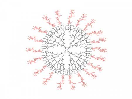 |
| A carefully constructed spherical nanoparticle coated in sialic acid lures the influenza A virus to its doom. (Image: Rensselaer Polytechnic Institute)
|
|
“Instead of blocking the virus, we mimicked its target – it’s a completely novel approach,” said Robert Linhardt, a glycoprotein expert and Rensselaer Polytechnic Institute professor who led the research. “It is effective with influenza and we have reason to believe it will function with many other viruses. This could be a therapeutic in cases where vaccine is not an option, such as exposure to an unanticipated strain, or with immune-compromised patients.”
|
|
The project is a collaboration between researchers within the Center for Biotechnology and Interdisciplinary Studies (CBIS) at Rensselaer and several institutions in South Korea including Kyungpook National University. Lead author Seok-Joon Kwon, a CBIS research scientist, coordinated the project across borders, enabling the South Korean institutions to test a drug designed and characterized at Rensselaer. Authors included Kwon, Linhardt, Ravi S. Kane, Jonathan S. Dordick, Marc Douaisi, and Fuming Zhang at Rensselaer; and Korean researchers Kyung Bok Lee, Dong Hee Na, Jong Hwan Kwak, Eun Ji Park, Jong-Hwan Park, Hana Youn, and Chang-Seon Song.
|
|
To access the interior of a cell and replicate itself, influenza A must first bind to the cell surface, and then cut itself free. It binds with the protein hemagglutinin, and severs that tie with the enzyme neuraminidase. Influenza A produces numerous variations each of hemagglutinin and neuraminidase, all of which are antigens within the pathogen that provoke an immune system response. Strains of influenza A are characterized according to the variation of hemagglutinin and neuraminidase they carry, thus the origin of the familiar H1N1 or H3N2 designations.
|
|
Medications to counter the virus do exist, but all are vulnerable to the continual antigenic evolution of the virus. A yearly vaccine is effective only if it matches the strain of virus that infects the body. And the virus has shown an ability to develop resistance to a class of therapeutics based on neuraminidase inhibitors, which bind to and block neuraminidase.
|
|
The new solution targets an aspect of infection that does not change: all hemagglutinin varieties of influenza A must bind to human sialic acid. To trap the virus, the team designed a dendrimer, a spherical nanoparticle with treelike branches emanating from its core. On the outermost branches, they attached molecules, or “ligands,” of sialic acid.
|
|
The research found that the size of the dendrimer and the spacing between the ligands is integral to the function of the nanoparticle. Hemagglutinin occurs in clusters of three, or “trimers,” on the surface of the virus, and researchers found that a spacing of 3 nanometers between ligands resulted in the strongest binding to the trimers. Once bound to the densely packed dendrimer, viral neuraminidase is unable to sever the link. The coat of the virus contains millions of trimers, but the research revealed that only a few links provokes the virus to discharge its genetic cargo and ultimately self-destruct.
|
|
A different approach, using a less structured nanoparticle, had been previously tested in unrelated research, but the nanoparticle selected proved both toxic, and could be inactivated by neuraminidase. The new approach is far more promising.
|
|
“The major accomplishment was in designing an architecture that is optimized to bind so tightly to the hemagglutinin, the neuraminidase can’t squeeze in and free the virus,” said Linhardt. “It’s trapped.”
|