| Feb 13, 2006 |
Counting Individual Biomolecules Using Color-Coded Nanoparticles
|
|
(Nanowerk News) Using two brightly colored fluorescent nanoparticles, a team of investigators led by Shuming Nie, Ph.D., of Emory University and Georgia Institute of Technology, has developed a method of counting single biomolecules as they flow through the channels of a microfluidics device. With additional work, this new approach to molecular detection could lead to earlier diagnosis of cancer and provide researchers with a versatile tool for studying single molecule processes inside living cells.
|
|
Reporting their work in the Jan. 19, 2006 edition of Analytical Chemistry, "Counting Single Native Biomolecules and Intact Viruses with Color-Coded Nanoparticles", Nie and his colleagues describe how they use coincidental detection of both red and green quantum dots to detect individual biomolecules and virus particles. Each of the two colors of quantum dots is labeled with a molecule that can bind to a different segment of the biomolecule they wish to detect. For detecting proteins and virus particles, the researchers use two different monoclonal antibodies, each of which will bind to a different portion of the protein or virus in question. To detect a particular gene, the investigators use two short nucleic acids, each of which binds to a specific region of the gene’s DNA sequence. The antibodies and short nucleic acids are known as "capture probes".
|
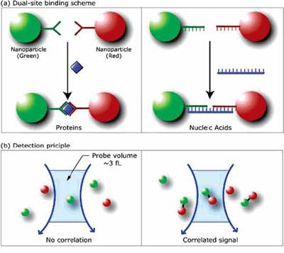 |
In the top figure, quantum dots of two different colors bind to different regions of the same biomolecule. In the bottom figure, unbound quantum dots flowing past a detector (left) do not signal that the biomolecule is present. Only when both colors of quantum dots pass the detector simultaneously is the biomolecule present. (Courtesy of Shuming Nie, Ph.D., Emory University and Georgia Institute of Technology.)
|
|
When these modified quantum dots are added to a biological sample, the capture probes bind to any of their respective targets present in the sample (Figure 1a). As the sample flows past an optical detector in a microfluidics device, the mixture is irradiated with a single frequency of ultraviolet that will trigger fluorescence by both of the red and green quantum dots. Only when the detector senses both colors flowing past it at the exact same time does it signal that the molecules in question are present in the sample (Figure 1b). This "coincident detection" method greatly reduces the background signal that results from nonspecific binding and eliminates the need to separate any unbound quantum dots prior to detection.
|
|
The researchers note that though they used two colors of quantum dots in the current work, their technique could be extended to multiple pairs of colored nanoparticles, permitting the detection of several diagnostic biomolecules in the same sample. The investigators estimate this technique should be capable of analyzing as many as 10 different biomolecules in a single assay. If so, this device could be an early prototype of a molecular screening device for determining cancer risk.
|