| Jun 02, 2016 |
Red for go, green for done
|
|
(Nanowerk News) A multifunctional molecular probe that can destroy cancer cells has been designed by A*STAR researchers. Unlike other probes that monitor only a single process, the new probe emits red and green light while killing cells, allowing surgeons to monitor its activation and assess the response of cancer cells to therapy (Advanced Functional Materials, "Light-Up Probe for Targeted and Activatable Photodynamic Therapy with Real-Time In Situ Reporting of Sensitizer Activation and Therapeutic Responses").
|
|
Molecular probes have been developed for light-excited treatment of tumors, but until now such probes indicate either drug activation or therapeutic response, not both. Bin Liu of the A*STAR Institute of Materials Research and Engineering and her collaborator Ben Zhong Tang from the Hong Kong University of Science and Technology have developed a probe that allows both processes to be monitored.
|
 |
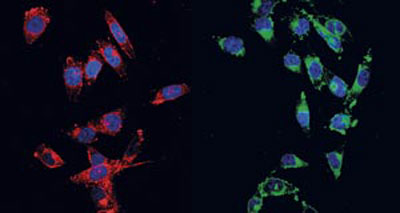
| The probe developed in this study emits red light (left) on reacting with an antioxidant in a cell, indicating that it is ready to be irradiated with the excitation light. After excitation, it emits green light (right), indicating that the cell is in its death throes. (© Wiley)
|
|
“It was very challenging to design a single probe that can monitor both processes and with the same excitation light,” says Liu.
|
|
The probe contains two compounds, one which emits green light, the other, red. These compounds only emit light when they aggregate, meaning the probe emits light only when these molecules are released from it and crowd together.
|
|
The probe employs quite an elaborate strategy to destroy cancer cells. It gains entry by latching on to a receptor on the cell membrane, then when engulfed in a pocket, it is able to pass through the membrane.
|
|
Once inside the cell, it reacts with glutathione, an antioxidant already present, causing it to simultaneously release a sensor probe and produce a ‘photosensitizer’, which fluoresces red.
|
|
When the photosensitizer is excited by light, it generates oxygen radicals that induce cell death. At the same time, an enzyme is activated and it reacts with the sensor probe which then fluoresces green.
|
|
Thus, the red fluorescence can be used to optimize the placement and timing of the excitation light for photodynamic therapy, while the green fluorescence can be used to image the response to the therapy.
|
|
“Our work highlights the feasibility of developing a simple, small-molecule probe, which shows activated therapeutic effect and in situ reporting of the therapeutic response at the same time,” says Liu. “This smart probe could help to forecast specific therapeutic regimes for individuals to realize the ultimate goal of personalized medicine.”
|
|
The team plans to play with the design of the molecule to realize excitation and emission at longer wavelengths. This will allow image-guided therapy deeper within the body as longer wavelengths penetrate further into tissue.
|

