DNA Amplification: Definition, PCR, Isothermal Methods, and Uses
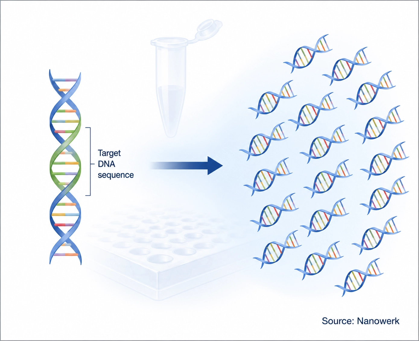
Definition: DNA amplification is the laboratory process of making many copies of a selected DNA sequence from a very small starting amount. It allows DNA that would otherwise be too scarce to detect to be measured, sequenced, cloned, or analyzed. The polymerase chain reaction (PCR) is the best-known amplification method, but isothermal and whole-genome techniques can amplify DNA without using the same temperature-cycling approach.
What Is DNA Amplification?
DNA amplification turns a small amount of genetic material into a workable signal. A sample may contain only a few molecules of the target sequence: a trace of DNA from a forensic stain, a small number of viral genomes in a swab, DNA from a single cell, or a fragment recovered from an ancient specimen. Most analytical methods cannot reliably read such tiny amounts directly, so amplification raises the copy number until the sequence can be detected and used.
At a glance:
- Goal: make many copies of a chosen DNA sequence from a small starting amount
- Best-known method: the polymerase chain reaction (PCR)
- Core ingredients: template DNA, primers, DNA polymerase, nucleotides, buffer, and magnesium ions
- Typical gain: millions to billions of copies under efficient conditions
- Alternatives: isothermal amplification methods such as LAMP and RPA, plus whole-genome amplification
- Main uses: diagnostics, sequencing, cloning, forensics, biosensing, and research
Amplification is best understood as a goal rather than a single technique. PCR reaches that goal through repeated heating and cooling. Isothermal methods reach it at one fixed temperature using enzymes that separate DNA strands without thermal cycling. Whole-genome amplification attempts to copy DNA across an entire genome rather than focusing on one short target. All of these approaches rely on the same central idea: a DNA polymerase reads a template strand and builds a complementary strand from nucleotides.
Key distinction: DNA amplification is the general process; PCR is one method; qPCR, RT-PCR, digital PCR, LAMP, RPA, and whole-genome amplification are specialized versions or alternatives. All PCR is DNA amplification, but not all DNA amplification is PCR.
How Does DNA Amplification Work?
Every DNA amplification method depends on a template, primers, nucleotides, and a DNA polymerase. The template is the DNA sequence to be copied. The primers are short oligonucleotides that bind to matching sequences on the template and define the region to be amplified. The polymerase is an enzyme that extends the primers by adding free nucleotides, producing new DNA strands that can become templates in later rounds.
In PCR, the reaction repeats three temperature steps. First, the sample is heated to about 95°C to separate the double helix into single strands, a step called denaturation. The temperature is then lowered, typically to about 50–65°C, so the primers can anneal to their matching sequences. Finally, the reaction is warmed to around 72°C, where a heat-stable polymerase extends each primer and copies the target. Repeating these steps 20–40 times can increase a target sequence by orders of magnitude.
PCR became practical because heat-stable polymerases can survive repeated high-temperature denaturation. The classic example is Taq polymerase from the heat-loving bacterium Thermus aquaticus, whose use in PCR was reported in 1988. PCR as a technique was conceived by Kary Mullis in the early 1980s, work for which he shared the 1993 Nobel Prize in Chemistry.
Primer design is often the most important design decision in an amplification assay. Primers determine where copying starts and ends, so they largely determine specificity. Poorly designed primers can bind to the wrong sequence, form primer dimers, or amplify background DNA instead of the intended target.
The PCR Toolkit: Conventional PCR, qPCR, RT-PCR, and Digital PCR
Conventional, or end-point, PCR amplifies a target and analyzes the product after the reaction is complete, often by gel electrophoresis. It is commonly used to test whether a target is present, check the size of a DNA fragment, prepare inserts for cloning, support genotyping, or recover DNA for downstream work. It is simple and robust, but it provides limited quantitative information.
Quantitative PCR (qPCR), also called real-time PCR, measures product formation during the reaction using fluorescence. The earlier the fluorescent signal crosses a threshold, the more target was present at the start. qPCR can support relative quantification, such as comparing gene expression between samples, or standard-curve-based absolute quantification. qPCR and RT-qPCR are workhorses of gene expression measurement and pathogen load testing, and their reliability depends on careful assay design, controls, and reporting standards such as the MIQE guidelines.
When the starting molecule is RNA, it must first be converted into complementary DNA (cDNA) by reverse transcriptase. The cDNA is then amplified by PCR or qPCR. This combined workflow is called RT-PCR or RT-qPCR and is widely used for RNA viruses, mRNA analysis, and gene expression studies.
Digital PCR (dPCR) partitions a sample into thousands or millions of small reactions, often droplets or microchambers. After amplification, each partition is scored as positive or negative, and the starting molecule count is calculated statistically from the fraction of negative partitions. Because it counts molecules after partitioning rather than relying mainly on a calibration curve, dPCR is valuable for rare-mutation detection, copy-number analysis, liquid-biopsy biomarkers, and samples that contain PCR inhibitors.
Isothermal Amplification: Copying DNA Without a Thermocycler
PCR traditionally ties amplification to a thermocycler, which adds cost, power requirements, and instrumentation complexity. Isothermal amplification methods reduce that dependency by running at one constant temperature. Instead of using heat to separate DNA strands again and again, they use specialized enzymes and primer designs to make the template accessible to polymerase.
Loop-mediated isothermal amplification (LAMP) is one of the most widely used isothermal methods. It uses a strand-displacing polymerase and several primers that recognize multiple regions of the target sequence. LAMP usually runs at about 60–65°C and can produce large amounts of DNA quickly, often with a visible color, fluorescence, or turbidity readout. It is attractive for point-of-care pathogen testing, but primer design is more complex than in PCR.
Recombinase polymerase amplification (RPA) operates at lower temperatures, often around 37–42°C. A recombinase helps primers invade double-stranded DNA, reducing the need for precise heating. This makes RPA useful for rapid and field-oriented assays, although practical testing still depends on sample preparation, contamination control, and reliable readout chemistry.
LAMP and RPA are often used qualitatively – for example, to ask whether a pathogen target is present – but real-time fluorescence and other monitored formats can make them semi-quantitative or quantitative. Their main advantage is practical rather than magical: they shift amplification from instrument-heavy laboratory workflows toward simpler heaters, portable devices, and integrated diagnostic platforms. Amplification is also central to many nanobiotechnology sensors, where nanoscale probes, particles, or surfaces detect amplified nucleic acid signals.
Whole-Genome Amplification
Most amplification assays target one defined DNA region. Whole-genome amplification (WGA) has a different aim: it attempts to amplify DNA across the genome from a very small starting amount, such as a single cell or a few degraded fragments. This is important in single-cell genomics, preimplantation genetic testing, microbial genomics, and forensic analysis.
The central limitation of WGA is uneven coverage. Some regions amplify better than others, and missing or overrepresented regions can affect sequencing and variant calling. Multiple displacement amplification, degenerate-oligonucleotide primed PCR, and other WGA methods each balance coverage, fidelity, yield, and bias differently. For this reason, WGA is powerful, but it is not simply a scaled-up version of target-specific PCR.
Comparison of Common DNA Amplification Methods
| Method | Temperature strategy | Quantitative? | Equipment burden | Common use |
|---|---|---|---|---|
| Conventional PCR | Repeated thermal cycles | Limited; usually end-point | Thermocycler | Target detection, cloning, genotyping |
| qPCR / real-time PCR | Repeated thermal cycles | Yes; relative or standard-curve based | Real-time thermocycler | Gene expression, pathogen load, copy number |
| RT-PCR / RT-qPCR | Reverse transcription plus PCR or qPCR | Depends on format | Thermocycler or real-time thermocycler | RNA viruses, mRNA analysis |
| Digital PCR | Thermal cycling in partitions | Yes; absolute molecule count after partitioning | Partitioning system and reader | Rare mutations, liquid biopsy, copy-number analysis |
| LAMP | Constant ~60–65°C | Often qualitative; real-time formats possible | Simple heater or integrated device | Point-of-care pathogen detection |
| RPA | Constant ~37–42°C | Often qualitative; real-time formats possible | Low-temperature heater; sometimes minimal equipment | Field and rapid diagnostics |
| Whole-genome amplification | Method-dependent; often isothermal | Genome-wide rather than target-specific | Standard molecular biology workflow | Single-cell genomics, low-input sequencing |
The best method depends on the question. Digital PCR gives precise molecule counts but requires partitioning hardware. LAMP and RPA simplify instrumentation but can be harder to design and control. qPCR sits in the middle and remains the default for many quantitative assays. In practice, laboratories choose the technique according to sample type, target abundance, required precision, turnaround time, and testing environment.
What Is DNA Amplification Used For?
DNA amplification is a foundational step in modern genetics and biotechnology. In clinical diagnostics, it helps detect pathogens, quantify microbial or viral load, identify cancer-associated mutations, and support prenatal, newborn, and inherited-disease testing. In molecular cloning and recombinant DNA work, PCR generates specific fragments for engineering and analysis.
Amplification also supports sequencing. Many next-generation sequencing workflows amplify library molecules before or during sequencing so that each fragment produces a detectable signal. Beyond the clinic and the sequencer, amplification enables forensic DNA profiling, food and species authentication, ancient DNA studies, environmental DNA surveys, antimicrobial-resistance monitoring, and genetic tests used in personalized medicine. Few molecular biology techniques connect basic research, diagnostics, sequencing, and forensic analysis as directly.
Limitations: Contamination, Bias, and Error
The extreme sensitivity that makes amplification powerful is also its main weakness. A single stray DNA molecule can be copied along with the intended target, creating a false positive. Carryover contamination from previous amplification products is especially serious, which is why amplification workflows use negative controls, physical separation of pre- and post-amplification work, clean reagents, and careful sample handling.
Amplification can also introduce error and bias. DNA polymerases occasionally make mistakes, so high-fidelity proofreading enzymes are used when exact sequence preservation matters. Some targets amplify more efficiently than others, which can distort quantitative comparisons and whole-genome data. Inhibitors in clinical, environmental, or forensic samples can suppress amplification and cause false negatives. Good assay design, controls, validated protocols, and appropriate reporting standards reduce these risks but do not eliminate them.
Frequently Asked Questions
What is the difference between DNA amplification and PCR? DNA amplification is the general process of making many copies of a DNA sequence, and PCR is the most widely used method for doing it. PCR uses repeated heating and cooling cycles, but other methods, including LAMP, RPA, and whole-genome amplification, also amplify DNA. All PCR is DNA amplification, but not all DNA amplification is PCR.
Is DNA amplification the same as DNA replication? DNA amplification and DNA replication both involve copying DNA, but they are not the same thing. DNA replication is the natural cellular process that copies an organism's genome before cell division. DNA amplification is a laboratory process that selectively increases the number of copies of a DNA target, often using PCR or a related method.
Why does DNA need to be amplified before analysis? Many samples contain too little target DNA to detect directly. A swab, blood sample, forensic trace, or ancient specimen may contain only a few copies of the sequence of interest. Amplification raises the copy number so the target can be measured, sequenced, cloned, or identified with confidence.
What is the difference between PCR and qPCR? Conventional PCR amplifies DNA and usually analyzes the product at the end of the reaction. qPCR, or quantitative PCR, monitors product formation during each cycle using fluorescence. This allows the starting amount of target DNA or cDNA to be estimated, either relatively or by comparison with a standard curve.
Can DNA be amplified without a thermocycler? Yes. Isothermal amplification methods run at a single temperature and do not require the repeated heating and cooling used in PCR. Methods such as LAMP and RPA use specialized enzymes to separate and copy DNA strands, which makes them useful for point-of-care and field testing.
How is RNA amplified if amplification copies DNA? RNA is first converted into complementary DNA, or cDNA, by reverse transcriptase. The cDNA is then amplified by PCR or another DNA amplification method. This combined workflow is called RT-PCR or RT-qPCR and is widely used for RNA viruses and gene expression studies.
Further Reading
Clinical Chemistry, MIQE 2.0: Revision of the Minimum Information for Publication of Quantitative Real-Time PCR Experiments Guidelines
Sensors, dPCR: A Technology Review
Frontiers in Microbiology, Advancements and Applications of Loop-Mediated Isothermal Amplification Technology: A Comprehensive Overview
