DNA Methylation – Epigenetic Marks in Gene Regulation, Development, and Disease
What Is DNA Methylation?
DNA methylation is an epigenetic modification in which a methyl group is added to a DNA base without changing the underlying DNA sequence. In mammals, the best-known form is 5-methylcytosine (5mC), produced when a methyl group is attached to the carbon-5 position of cytosine, most often at CpG dinucleotides.
In brief DNA methylation is a reversible chemical mark on DNA that helps cells regulate genes, preserve cell identity, silence repetitive elements, and record developmental or environmental history. Its meaning depends on where it occurs in the genome.
DNA methylation is one of the main ways cells create stable gene-regulation programs on top of the genetic code. The same genome can support a neuron, liver cell, immune cell, or stem cell because each cell type maintains a different pattern of accessible, silent, poised, and tissue-specific regulatory regions. These patterns are part of the epigenome.
In a typical mammalian somatic cell, most isolated CpG sites are methylated, while many CpG-dense promoter regions, called CpG islands, remain unmethylated. Non-CpG methylation also occurs in mammals and is especially notable in pluripotent cells and neurons. This distribution is important: methylation across repetitive DNA and transposable elements helps protect genome stability, whereas abnormal methylation of CpG-island promoters can silence genes that should remain active.
The effect of methylation depends strongly on genomic context. Promoter methylation is usually associated with transcriptional repression, gene-body methylation is often found in actively transcribed genes, and enhancer methylation can mark regulatory regions as inactive or lineage-restricted. DNA methylation should therefore not be reduced to a simple on/off switch.
Synonyms and related terms: cytosine methylation, CpG methylation, 5-methylcytosine, 5mC, DNA methylome. The complete map of methylation sites across a genome is called the methylome.
Not to be confused with: DNA hydroxymethylation, histone methylation, RNA methylation, or genetic mutation. DNA methylation changes a chemical mark on DNA; it does not change the A, C, G, and T sequence itself.
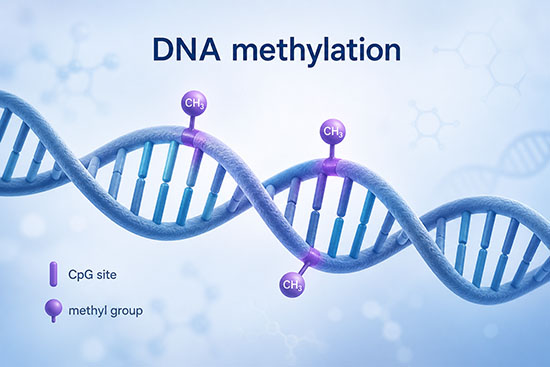
DNA methylation adds methyl groups to DNA bases, most often cytosines at CpG sites in mammals, changing how genes and regulatory regions are interpreted without altering the genetic sequence. (Image: Nanowerk)
How DNA Methylation Fits Into Epigenetics
DNA methylation is one layer of epigenetic regulation. It works together with histone modifications, chromatin accessibility, non-coding RNAs, and three-dimensional genome organization to control which parts of the genome are active, silent, poised, or insulated from nearby regulatory signals.
| Epigenetic layer | What changes | Typical role |
|---|---|---|
| DNA methylation | Chemical marks on DNA bases, especially cytosine | Stable gene regulation, repeat silencing, imprinting, and cell-state memory |
| Histone modifications | Chemical marks on histone proteins | Open, compact, activate, or repress chromatin regions |
| Chromatin accessibility | How exposed DNA is to regulatory proteins | Controls whether transcription factors can bind regulatory DNA |
| Non-coding RNAs | RNA molecules that do not encode proteins | Guide chromatin regulators, silence regions, or modulate gene expression |
| 3D genome organization | Physical folding of chromosomes | Brings enhancers, promoters, insulators, and domains into functional contact |
Why DNA Methylation Matters
DNA methylation matters because it connects genome sequence, chromatin state, cell identity, development, disease risk, and biotechnology. It is stable enough to preserve cell-state memory through many rounds of cell division, but dynamic enough to be remodeled during embryogenesis, germ-cell formation, immune activation, aging, and disease.
For biotechnology, methylation is useful for three main reasons. First, it is mechanistic: methylation can contribute to gene silencing, transposon repression, imprinting, and regulatory-region activity. Second, it is measurable: methylation can be mapped at single-base resolution or profiled at population scale. Third, it is clinically informative: disease states often leave characteristic methylation signatures in tissues and cell-free DNA.
Where Methylation Occurs in the Genome
| Genomic context | Typical methylation pattern | Functional meaning |
|---|---|---|
| CpG-island promoters | Often unmethylated in normal active genes | Methylation is usually associated with stable repression, although it may be a cause, reinforcement, or consequence of silencing depending on context |
| Gene bodies | Often methylated in expressed genes | Associated with transcriptional elongation, splicing patterns, and suppression of cryptic promoters |
| Enhancers | Often low methylation when active, higher methylation when inactive | Helps distinguish active, poised, and lineage-restricted regulatory elements |
| Repetitive DNA and transposons | Usually heavily methylated | Suppresses mobile elements and protects genome integrity |
| Imprinting control regions | Parent-of-origin-specific methylation | Allows expression from only the maternal or paternal allele for selected genes |
Plants and fungi use additional methylation contexts beyond mammalian CpG methylation. Plants methylate cytosines in CG, CHG, and CHH sequence contexts, where H is A, C, or T. Bacteria also methylate DNA, often on adenine or cytosine bases, but bacterial methylation is usually discussed in the context of restriction-modification systems, DNA replication, and gene regulation rather than mammalian epigenetics.
How DNA Methylation Works
The methyl group used for DNA methylation comes from S-adenosylmethionine (SAM), a universal cellular methyl donor. DNA methyltransferase enzymes, abbreviated DNMTs, catalyze the reaction by flipping the target cytosine out of the DNA helix and transferring a methyl group to its 5-position, producing 5-methylcytosine.
Mammalian cells use two broad methylation modes. De novo methylation establishes new methylation patterns, especially during early development and germ-cell maturation. Maintenance methylation copies existing patterns after DNA replication. When a methylated CpG is replicated, the parental strand remains methylated while the new strand is initially unmethylated. DNMT1, guided by UHRF1 and other chromatin cues, methylates the new strand so daughter cells inherit the parental pattern.
Methylation can repress gene expression in several ways. A methylated promoter may directly block transcription-factor binding, or it may recruit methyl-CpG-binding proteins that bring in chromatin-remodeling and histone-modifying complexes. The result is often denser chromatin and reduced access by the transcription machinery.
Writers, Readers, and Erasers
DNA methylation is often described through a writer-reader-eraser framework. Writers add the mark, readers interpret it, and erasers remove or modify it. This framework is useful, but simplified: methylation state is also shaped by transcription factors, histone marks, nucleosome positioning, replication timing, metabolism, and DNA repair.
| Component | Main examples | Role | Why it matters |
|---|---|---|---|
| Writers | DNMT1, DNMT3A, DNMT3B; DNMT3L as a catalytically inactive cofactor | Add methyl groups to cytosine | Establish and maintain methylation landscapes during development and cell division |
| Readers | MeCP2, MBD1, MBD2, MBD4, UHRF1, Kaiso-family proteins | Recognize methylated DNA or hemimethylated DNA | Link methylation to chromatin compaction, transcriptional repression, repair, and maintenance copying |
| Erasers and converters | TET1, TET2, TET3, base-excision repair enzymes | Oxidize 5mC to 5hmC, 5fC, and 5caC; repair pathways can restore unmodified cytosine | Enable active demethylation and create oxidized cytosine marks with regulatory roles |
The DNMT family has clinically important specialization. DNMT1 is the main maintenance methyltransferase. DNMT3A and DNMT3B establish new methylation patterns. DNMT3L lacks catalytic activity but stimulates DNMT3A and DNMT3B, especially in the germ line. DNMT3C is a rodent-specific paralog involved in methylating young transposons in male germ cells.
TET enzymes changed how scientists think about methylation. DNA methylation was once viewed mainly as a stable, one-way repressive mark. TET1, TET2, and TET3 can oxidize 5mC to 5-hydroxymethylcytosine (5hmC), then to 5-formylcytosine and 5-carboxylcytosine. These oxidized bases can be diluted through replication or removed through repair, creating routes for passive and active demethylation.
5hmC is not merely an intermediate on the way back to unmodified cytosine. It is enriched in neurons, embryonic stem cells, active enhancers, and gene bodies, and it often marks regulatory regions that are transcriptionally active or developmentally flexible.
Roles in Development and Genome Regulation
DNA methylation is extensively reprogrammed during mammalian development. After fertilization, much of the parental methylation landscape is erased, allowing the early embryo to reset developmental potential. As cells commit to specific lineages, DNMT3A and DNMT3B help rebuild methylation patterns that support tissue-specific gene regulation. A second major reprogramming event occurs in primordial germ cells so the next generation can establish sex-specific and parent-of-origin-specific marks.
Genomic imprinting depends on methylation marks inherited from either the mother or the father. These marks cause selected genes to be expressed from only one parental allele. Errors in imprinting are associated with disorders such as Prader-Willi syndrome, Angelman syndrome, and Beckwith-Wiedemann syndrome.
DNA methylation also contributes to X-chromosome inactivation in female mammals. X inactivation is initiated by the non-coding RNA XIST and reinforced by chromatin changes, including methylation of promoters on the inactive X chromosome. The result is dosage compensation: balancing expression of X-linked genes between XX and XY individuals.
Another core role is genome defense. Transposable elements and repetitive sequences make up a large fraction of mammalian genomes. DNA methylation, together with repressive histone marks, keeps many of these elements silent, reducing the risk of genome rearrangements, insertional mutagenesis, and inappropriate immune activation.
DNA Methylation in Cancer and Disease
Cancer cells often show a methylation paradox: widespread loss of methylation across repetitive and intergenic regions, combined with focal hypermethylation at selected promoters and CpG islands. Global hypomethylation can contribute to chromosomal instability and transposon activation. Focal hypermethylation can silence tumor-suppressor and DNA-repair genes, such as MLH1 in some colorectal cancers and BRCA1 in some breast and ovarian cancers.
Mutations in methylation regulators can also drive disease. DNMT3A mutations are common in clonal hematopoiesis and acute myeloid leukemia. TET2 loss is common in blood cancers and can reshape enhancer methylation. IDH1 and IDH2 mutations produce 2-hydroxyglutarate, an oncometabolite that inhibits TET enzymes and contributes to hypermethylated tumor states in glioma and leukemia.
Outside cancer, fragile X syndrome is a classic methylation-linked disorder. Expansion of CGG repeats near the FMR1 gene triggers promoter hypermethylation and gene silencing, reducing production of the FMRP protein needed for normal synaptic function. Mutations in MECP2, a methylated-DNA reader, cause Rett syndrome, showing that interpreting methylation marks is as important as writing them.
Many complex diseases show altered methylation patterns, including cardiovascular, autoimmune, metabolic, neurodegenerative, and psychiatric conditions. These associations require careful interpretation because methylation differences may contribute to disease, reflect altered cell composition, record exposure, or arise as a consequence of pathology.
How DNA Methylation Is Measured
Methylation can be measured globally, at selected loci, across panels of CpG sites, or genome-wide. The best method depends on the biological question, sample type, required resolution, DNA input, cost, and whether the experiment needs to distinguish 5mC from 5hmC.
| Method | What it measures | Strengths | Limitations |
|---|---|---|---|
| Bisulfite sequencing | Single-base methylation after chemical conversion of unmethylated cytosines | High resolution; compatible with targeted, reduced-representation, and whole-genome designs | Harsh chemistry damages DNA; standard bisulfite does not distinguish 5mC from 5hmC |
| Enzymatic methyl-seq | Single-base methylation using enzymatic conversion | Less DNA damage and often better library complexity | Newer workflows; cost and platform support vary |
| OxBS-seq, TAB-seq, TAPS, and related methods | 5mC and/or 5hmC with greater chemical specificity | Separates marks that standard bisulfite merges | More complex experimental design and analysis |
| Methylation arrays | Preselected CpG sites across the genome | Standardized, cost-effective, useful for cohorts and epigenetic clocks | Limited to probes on the array; less useful for non-model organisms or novel regions |
| Nanopore and other long-read sequencing | Native DNA modifications inferred directly from sequencing signals | Can combine methylation, haplotypes, repeats, and structural variants in long molecules | Calling accuracy, coverage, and analysis pipelines must be carefully validated |
| Methylation-specific PCR and digital PCR | Selected methylated loci | Fast, sensitive, suitable for clinical assays and low-input samples | Targeted only; requires prior knowledge of the marker |
In many datasets, methylation at a CpG site is reported as a percentage or beta value, ranging from 0 for unmethylated to 1 for fully methylated. In bulk tissue, this value often reflects a mixture of cells, alleles, and DNA fragments rather than a single uniform state.
Bioinformatics is central to methylation analysis. Typical steps include quality control, alignment or signal processing, methylation calling, normalization, identification of differentially methylated positions or regions, annotation to genes and regulatory elements, and correction for confounders such as age, sex, ancestry, smoking, batch effects, and cell-type composition.
Applications in Biotechnology and Medicine
Cancer diagnostics and liquid biopsy
Tumors shed fragments of DNA into blood and other body fluids. Because cancer-specific methylation patterns can appear early and can reflect tissue of origin, methylation is a major signal for liquid-biopsy cancer detection, tumor classification, recurrence monitoring, and minimal residual disease assays. Some methylation assays are already used in specific clinical contexts, while broad multi-cancer early detection remains an evolving area where performance, follow-up pathways, reimbursement, and regulatory status differ by test and jurisdiction. A positive screening result is not a diagnosis; it must be followed by imaging, endoscopy, pathology, or other confirmatory testing.
Therapeutic demethylation
The nucleoside analogs azacitidine and decitabine are hypomethylating agents used in hematologic malignancies. They incorporate into nucleic acids and trap DNMT enzymes, leading to DNA hypomethylation and altered gene expression. Decitabine combined with cedazuridine enables oral exposure to decitabine for approved myelodysplastic syndrome and chronic myelomonocytic leukemia indications in the United States.
Epigenetic clocks and aging biology
DNA methylation patterns change reproducibly with age. Epigenetic clocks use selected CpG sites and statistical models to estimate chronological age, biological age, mortality risk, or pace of aging. These clocks are valuable research tools, but clock reversal should not automatically be interpreted as proven rejuvenation unless linked to meaningful clinical outcomes.
Cell identity, regenerative medicine, and quality control
Methylation profiles can verify cell type, detect incomplete reprogramming, identify culture drift, and flag aberrant differentiation in stem-cell and cell-therapy workflows. Because methylation captures cell-state memory, it can reveal differences that are not obvious from morphology or a small set of marker genes.
Environmental exposure and epidemiology
Smoking, diet, exercise, alcohol, stress, pollution, and other exposures can leave measurable methylation signatures. Some signatures are durable enough to reconstruct exposure history. Others are transient or confounded by changes in blood-cell composition, making causal interpretation difficult.
Targeted epigenome editing
Programmable epigenome editors can fuse catalytically inactive Cas9 or other DNA-binding modules to DNMT or TET domains. These systems allow researchers to add or remove methylation at selected loci without cutting DNA. They are powerful for testing whether a methylation change causes a gene-expression change, and they are being explored as potential therapeutics for durable gene silencing or reactivation.
Limitations and Common Pitfalls
A common pitfall is assuming that methylation always silences genes. This is broadly true for many promoter CpG islands, but not for all genomic contexts. Gene-body methylation, enhancer methylation, allele-specific methylation, and non-CpG methylation can have different meanings.
A second pitfall is confusing correlation with causation. Many methylation studies compare diseased and healthy samples, but the observed differences may be caused by altered cell mixtures, medication, inflammation, age, smoking, ancestry, or technical batch effects. Causal claims require perturbation experiments, longitudinal data, genetic instruments, or strong mechanistic support.
A third pitfall is ignoring cell-type composition. Blood, tumors, brain tissue, and organ biopsies contain mixtures of cells. A methylation difference may simply mean that one sample contains more immune cells, stromal cells, neurons, epithelial cells, or tumor cells than another. Deconvolution or purified cell populations are often needed.
A fourth pitfall is treating 5mC and 5hmC as the same mark. Standard bisulfite sequencing reads both as protected cytosines, so additional methods are needed when hydroxymethylation is biologically relevant, especially in brain, stem-cell, and cancer studies.
Typical Research Workflow
A DNA methylation project usually begins with a clear biological contrast: treated versus untreated cells, tumor versus matched normal tissue, young versus old samples, or responders versus non-responders. Researchers then choose a platform, define quality-control thresholds, account for cell composition and covariates, identify differentially methylated sites or regions, and connect those regions to genes, enhancers, pathways, or clinical outcomes.
The strongest studies validate findings in an independent cohort and, when possible, test function directly. For example, a promoter found to be hypermethylated in tumors can be tested by demethylating the region, restoring gene expression, and measuring effects on cell growth, differentiation, drug response, or tumor formation.
Key Terms Related to DNA Methylation
| Term | Meaning |
|---|---|
| 5mC | 5-methylcytosine, the main methylated cytosine mark in mammalian DNA |
| 5hmC | 5-hydroxymethylcytosine, an oxidized cytosine mark produced by TET enzymes |
| CpG island | A CpG-rich genomic region, often overlapping a gene promoter |
| DNMT | DNA methyltransferase, an enzyme that adds methyl groups to DNA |
| TET enzyme | An enzyme that oxidizes 5mC and participates in active or passive demethylation |
| Methylome | The genome-wide pattern of DNA methylation |
| Differentially methylated region | A genomic region whose methylation differs between samples or conditions |
| Imprinting | Parent-of-origin-specific gene expression controlled partly by methylation |
| Non-CpG methylation | Cytosine methylation outside CpG sites, especially notable in pluripotent cells and neurons |
| Epigenetic clock | A model that estimates age-related biology from DNA methylation patterns |
Frequently Asked Questions
Is DNA methylation reversible? Yes. Methylation can be lost passively when DNA replicates without maintenance methylation, and it can be removed actively through TET-mediated oxidation followed by repair or dilution. Some methylation states are highly stable, while others are dynamic.
Does DNA methylation always silence genes? No. Promoter CpG-island methylation usually represses transcription, but gene-body methylation is often associated with active transcription, and methylation at enhancers or repeats has context-specific effects.
Is DNA methylation inherited? Methylation patterns are inherited through cell divisions within the body, which helps maintain cell identity. Most methylation is reset between generations. Genomic imprinting is a well-established exception; broader environmentally driven transgenerational methylation inheritance in mammals is much harder to prove and remains context-dependent.
Can lifestyle or environment change DNA methylation? Yes. Smoking, diet, exercise, alcohol, stress, pollution, and other exposures can leave methylation signatures. However, the presence of a methylation signature does not automatically prove that the exposure caused disease or that reversing the signature will improve health.
How is DNA methylation different from a genetic mutation? A mutation changes the DNA sequence. DNA methylation changes a chemical mark on the DNA. Methylation can influence gene expression and can be copied through cell division, but it is generally more reversible than sequence mutation.
Can methylation tests diagnose cancer? Some methylation assays are used clinically, and many more are being developed. Screening or liquid-biopsy tests can indicate a cancer-associated signal, but positive results require clinical follow-up and confirmation. Performance depends on cancer type, stage, assay design, and intended use.
Selected References
Smith Z.D., Hetzel S., Meissner A. “DNA methylation in mammalian development and disease.” Nature Reviews Genetics 26, 7–30 (2025). DOI: 10.1038/s41576-024-00760-8
Wu X., Zhang Y. “TET-mediated active DNA demethylation: mechanism, function and beyond.” Nature Reviews Genetics 18, 517–534 (2017). DOI: 10.1038/nrg.2017.33
Horvath S., Raj K. “DNA methylation-based biomarkers and the epigenetic clock theory of ageing.” Nature Reviews Genetics 19, 371–384 (2018). DOI: 10.1038/s41576-018-0004-3
Pharo H.D., Vedeld H.M., Sjurgard I.V., Pinto R., et al. “From concept to clinic: a roadmap for DNA methylation biomarkers in liquid biopsies.” Oncogene 44, 4814–4831 (2025). DOI: 10.1038/s41388-025-03624-5
Simpson J.T., Workman R.E., Zuzarte P.C., et al. “Detecting DNA cytosine methylation using nanopore sequencing.” Nature Methods 14, 407–410 (2017). DOI: 10.1038/nmeth.4184