| Posted: Sep 20, 2017 |
New microscope technology provides a detailed look at structure and composition of materials
(Nanowerk News) At their core, electron microscopes work a lot like a movie projectors. A high-powered beam passes through a material and it projects something — usually something we really want to see — onto a screen on the other side. With most electron microscopes, however, capturing data is like trying to project a movie onto a dirty screen that is too small to see the whole projection. But a new camera technology, tested by researchers at Drexel University, is enabling the microscopes to present a clearer, more complete and detailed look at their featured presentation.
|
|
Using a direct detection camera and an image filter, the group discovered that it can obtain a crisper picture of chemical structure and composition, and to obtain this data quite rapidly. It is also sensitive enough to operate the microscope in a way that allows scientists to study fragile, biological samples without damaging them. Drexel is the first to combine the use of these technologies to give researchers a detailed, clear look at the mechanisms behind chemical and physical reactions almost as quickly as they occur.
|
|
The team, led by Mitra Taheri, PhD, Hoeganaes associate professor in Drexel’s College of Engineering and director of the Dynamic Characterization Group in the Materials Science and Engineering Department, recently published its findings from a side-by-side test of a newly developed direct detection camera and a conventional indirect detection camera, both developed by Gatan. Their piece in the journal Nature Scientific Reports ("Direct Detection Electron Energy-Loss Spectroscopy: A Method to Push the Limits of Resolution and Sensitivity"), suggests that applying a direct detection sensor to standard electron energy-loss spectroscopy (EELS) will greatly enhance scientists ability to study a materials structure and chemistry at the nanometer level.
|
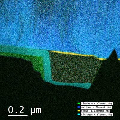 |
| Using a direct detection sensor in conjunction with electron-loss spectroscopy allows researchers to get a crisper look at the chemical composition and structure of materials. (click on image to enlarge)
|
|
“EELS is a popular technique that has been around for a while, however, the noise present in EELS is a major problem,” according to Jamie Hart, a doctoral researcher and coauthor of the paper. “By applying direct detection to EELS, we can greatly reduce the experimental noise, which will improve over real-time observation of dynamic processes, such as tracking the motion of lithium ions in Li-ion batteries, and it will aid the study of sensitive materials, like biological matter.”
|
|
In actuality any comparison between an electron microscope and a movie projector basically ends with the “on” button. Rather than pushing light through film, electron microscopes shoot a beam of charged electrons through the sample material being studied. They pass through the material and are recorded by a camera. The camera’s interpretation of the electrons’ journey can tell researchers a lot about the material. Some electrons pass through the material as if it wasn’t even there. Some pass through but change direction. Others pass through but are now moving at a different speed. All of these behaviors give researchers clues about the material’s chemical makeup and interior structure.
|
|
So adding a more sophisticated camera to a microscope can make a big difference in the data researchers can glean.
|
|
Taheri’s lab is the first to use this type of camera, a Gatan K2 direct detection camera, with an electron energy-loss spectroscopy (EELS) microscope — a type that draws its inferences about a sample by measuring how much energy electrons lose when they pass through it. EELS technology is typically used by researchers trying to determine which elements are present in a sample or the chemical structure of one given element. But by adding the direct detection camera to the system, Drexel’s team can Both determine which elements are present and understand each one’s chemical bonding.
|
|
“Direct detection provides higher energy resolution data, which helps us understand how atoms are bonded together, and it provides a greater energy field of view, allowing us to see more elements at once,” Hart said.
|
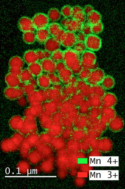 |
| Using a direct detection camera with an image filter, Drexel researchers are able to quickly and clearly obtain data about the chemical composition and structure of things like nanoparticles (pictured here).
|
|
The high sensitivity of the new camera means that it can probe a material more gently, exposing it to a lower dose of electrons, than other microscopes that blast a more powerful beam through the sample. Because of this, it can be used to study more fragile samples like viruses and bacteria.
|
|
“Using the low-noise direct detection sensor will basically reduce the number of electrons needed for analysis by a factor of 2-5, for biological samples which are easily destroyed under the electron beam, this makes a big difference. Also, if we want to watch some fast reaction, this lets us go to higher frame rates” Hart said.
|
|
To make it work, Gatan had to develop its own software interface with EELS and a protocol for operating it – which is no small task considering the device captures up to 1,600 frames per second, which equates to about 2 gigabytes of data, and runs so hot that it needs a constant circulation of water to keep it cool.
|
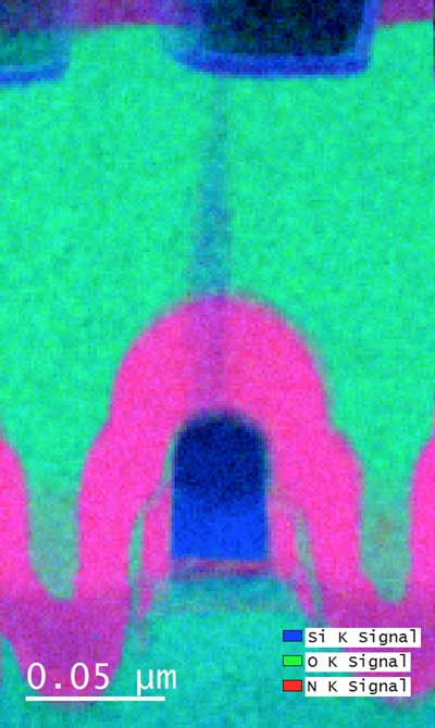 |
| Researchers will be able to use the direct detection technology with electron-loss spectroscopy to study a variety of materials, including biological samples like viruses and bacteria, and materials that are in development for computer components, energy storage and electromagnetic shielding.
|
|
“One of the greatest challenges with collecting high frame rate data is storage and processing. At the K2’s minimum, it is generating 400 images per second, each one is 16 million pixels, and that all adds up,” said Andrew Lang, a doctoral researcher in Taheri’s lab. “Our server rack can handle more than 3 gigabytes per second of data with some of the fastest solid state drive arrays available today.”
|
|
But all that effort is worth it, according to the team, when you can collect higher resolution, cleaner data in a shorter period of time than using a conventional camera. The team is currently using the K2 to examine materials that are in development for computer components, energy storage and electromagnetic shielding, and they suggest that it could also be used to study viruses and bacteria.
|


