| Nov 30, 2020 |
Raman holography
(Nanowerk News) Raman spectroscopy is widely used in analytical sciences to identify molecules via their structural fingerprint. In the biological context the Raman response provides a valuable label-free specific contrast that allows distinguishing different cellular and tissue contents.
|
|
Unfortunately, spontaneous Raman scattering is very weak, over ten orders of magnitude weaker than fluorescence. Unsurprisingly, fluorescence microscopy is often the preferred choice for applications such as live cell imaging.
|
|
Luckily, Raman can be enhanced dramatically on metal surfaces or in metallic nanogaps and this surface enhanced Raman scattering (SERS) can even overcome the fluorescence response.
|
|
Nanometric SERS probes are thus promising candidates for biological sensing applications, preserving the intrinsic molecular specificity.
|
|
Still, the effectiveness of SERS probes depends critically on the particle size, stability and brightness, and, so far, SERS-probe based imaging is rarely applied.
|
|
Now ICFO researchers Matz Liebel and Nicolas Pazos-Perez, working in the groups of ICREA professors Niek van Hulst (ICFO) and Ramon Alvarez-Puebla (Univ. Rovira i Virgili) have presented "holographic Raman microscopy".
|
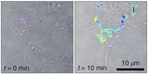 |
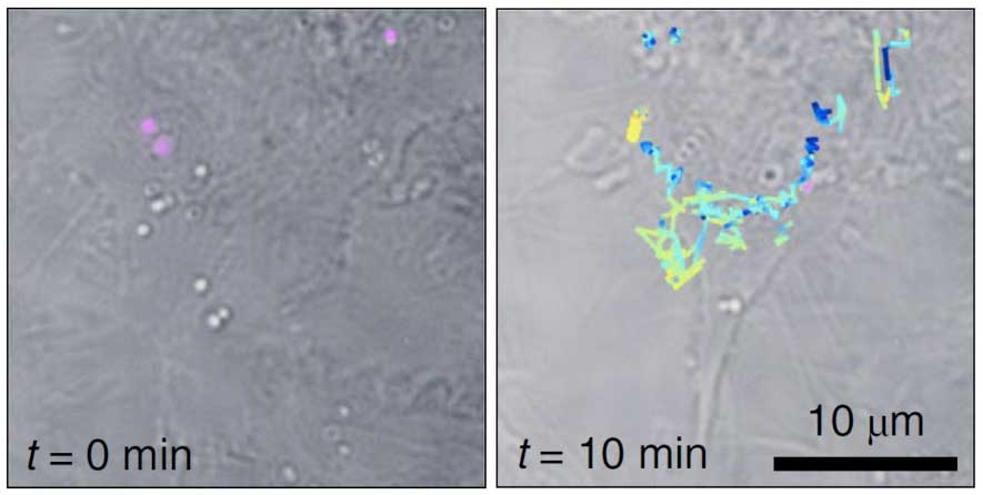
| Tracking of Live-cell SERS individual particles. The tracks of each of the particles are color coded to show the respective z-positions within the volume. (Image: ICFO/URV) (click on image to enlarge)
|
|
First, they synthesized plasmonic superclusters from small nanoparticle building blocks, to generate very strong electric fields in a restricted cluster size. These extremely bright SERS nanoprobes require very low illumination light exposure in the near-infrared, thus reducing potential photo-damage of live cells to a minimum, and allow wide-field Raman imaging.
|
|
Second, they took advantage of the bright SERS probes to realize 3D holographic imaging, using the scheme for incoherent holographic microscopy developed by Liebel and team in a study in Science Advances (Link).
|
|
Remarkably, the incoherent Raman scattering is made to "self-interfere" to achieve Raman holography for the first time.
|
|
Liebel and Pazos-Perez demonstrated Fourier transform Raman spectroscopy of the wide-field Raman images and were able to localize single-SERS-particles in 3D volumes from one single-shot. The authors then used these capabilities to identify and track single SERS nanoparticles inside living cells in three dimensions.
|
|
The results, published in Nature Nanotechnology ("Surface-enhanced Raman scattering holography") represent an important step towards multiplexed single-shot three-dimensional concentration mapping in many different scenarios, including live cell and tissue interrogation and possibly anti-counterfeiting applications.
|