| Aug 24, 2022 |
Researchers pioneer nanoelectronic sensor that simultaneously measures electrical and mechanical activity in heart cells(Nanowerk News) Using a suspended nanowire, a University of Massachusetts research team has, for the first time, created a tiny sensor that can simultaneously measure electrical and mechanical cellular responses in cardiac tissue, work promising for cardiac disease studies, drug testing and regenerative medicine. |
| Electrical and computer engineering (ECE) Ph.D. student Hongyan Gao, first author of the paper published by the journal Science Advances ("Bioinspired two-in-one nanotransistor sensor for the simultaneous measurements of electrical and mechanical cellular responses"), describes the invention as “a new tool for improved cardiac studies that has the potential for leading-edge applications in cardiac-disease experiments.” |
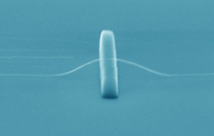 |
| SEM image of a suspended nanowire. (Image: Jun Yao) |
| Because the cell is a basic functional element in biology, its mechanical and electrical behaviors are two key properties that indicate cell state and consequently are important for health monitoring, disease diagnosis and tissue repair. |
| “A comprehensive assessment of cellular status requires knowledge of both mechanical and electrical properties at the same time,” says research team leader Jun Yao, ECE assistant professor and a biomedical engineering adjunct. These two properties are usually measured by different sensors, and the degree to which the cell’s function is disturbed increases with the number of sensors used. |
| The sensor is constructed from a 3D suspended semiconducting silicon nanowire. With its size much smaller than a single cell, the nanowire can tightly patch onto the cell membrane and “listen to” cellular activities very closely. It also has unique properties to convert “heard” bioelectrical and biomechanical activities into electrical sensing signals for detection. |
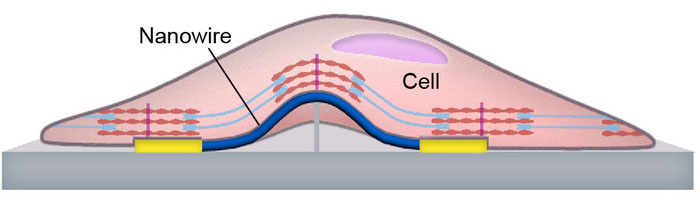 |
| Schematic of the sensor structure and cell-sensor interface. (Image: Jun Yao) |
| The research accomplishes goals proposed in a five-year, $500,000 grant from the National Science Foundation’s Faculty Early Career Development (CAREER) program that Yao received in 2019. |
| “Other than developing integrated biochips, our next step is to integrate the nanosensors on free-standing scaffolds to innervate in vitro tissue for deep-tissue studies,” Yao says. “In the long run, we hope the nanosensors can be safely delivered to living cardiac systems for improved health monitoring and early disease diagnosis.” |
| The concept of merging multiple sensing functions in one device will also broaden the capabilities of general bio-interface engineering, Yao says. |
| Source: University of Massachusetts Amherst |

