| Mar 27, 2023 |
Performing Bio-AFM on live cells
(Nanowerk News) A new application note discusses how atomic force microscope (AFM) systems can be used in various disciplines of biological research, such as imaging of live cells and bacteria, single-cell manipulation, or force spectroscopy on molecules, cells, or tissue.
|
|
To perform advanced biological experiments on cells, certain accessories are required. AFM becomes particularly powerful when combined with other techniques, especially with optical microscopy. The publisher of this appication note, Nanosurf, offers stages for all common brands of inverted microscopes that allow the AFM to be placed on top of the bio-sample, between the condenser and objective.
|
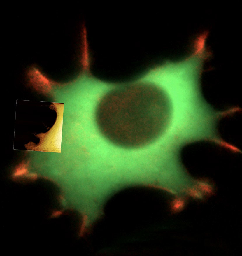 |
| Live fibroblast imaged with inverted light microscope and AFM in cell culture conditions. (Image: Nanosurf)
|
|
To establish physiological conditions, Nanosurf offers accessories like the AFM-compatible Live Cell Incubator that provides temperature, humidity, and CO2 control. For temperature-critical experiments, local heating via the Petri dish holder may not be sufficient, because a temperature gradient between the edge of the Petri dish and the center is difficult to prevent. To reduce temperature gradients in the Petri dish, a heating option that elevates the temperature around the complete optical microscope and AFM is available.
|
|
The Live Cell Incubator option seals the Petri dish, creating two chambers, while still allowing the cantilever holder to move freely. Combining the Live Cell Incubator with a suitable supply of humid gas, evaporation can be reduced to a minimum. Nanosurf also offers an advanced humidification scheme that allows controlling the humidity from ambient to 95% rel. hum. using a humidity sensor and microcontroller. The perfusion-insert option allows for fluid-exchange or fluid insertion without human intervention.
|
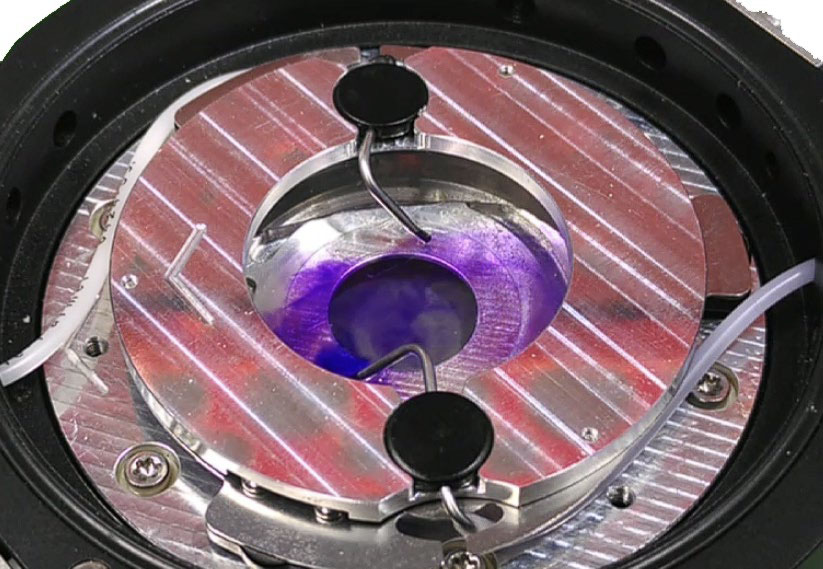 |
| Fluid inside the Petri dish can be added or removed using the perfusion insert. (Image: Nanosurf)
|
|
The 150 µm-stage is a Petri dish holder that has long-range piezo actuators integrated, allowing to precisely position and move the dish surface in height (z-axis) thus extending the z-range of the AFM. In single-cell force spectroscopy (SCFS), cell adhesion can be characterized down to the molecular level. The FluidFM probe combines the functionality of force-sensing cantilevers with that of micro-pipettes, offering a unique possibility of manipulating and probing cells. Using FuidFM probes that are micro-syringes instead of micropipettes extends FluidFM’s functionality even further.
|
|
In summary, Nanosurf offers a variety of accessories, in particular the different options for Petri dish holders, that enable researchers to perform advanced biological experiments on cells, maximize the potential of AFM in biological applications, and establish physiological conditions in terms of temperature, humidity, and CO2.
|
|
You can download the full white paper PDF here on the Nanosurf website.
|

