| Mar 19, 2024 |
Higher intensity x-ray beams don't always give stronger diffraction patterns(Nanowerk News) Turning up the intensity of x-ray beams used to probe the atomic structures of materials can actually reduce the intensity of x-rays scattered from the material, a RIKEN-led team has found. This rather counterintuitive result could be harnessed to produce ultrashort pulses of x-rays. |
| Scientists have been using x-rays to reveal the structure of crystalline materials for more than a century now. X-ray diffraction has played a large role in determining the structures of many key compounds, including DNA and cellulose. |
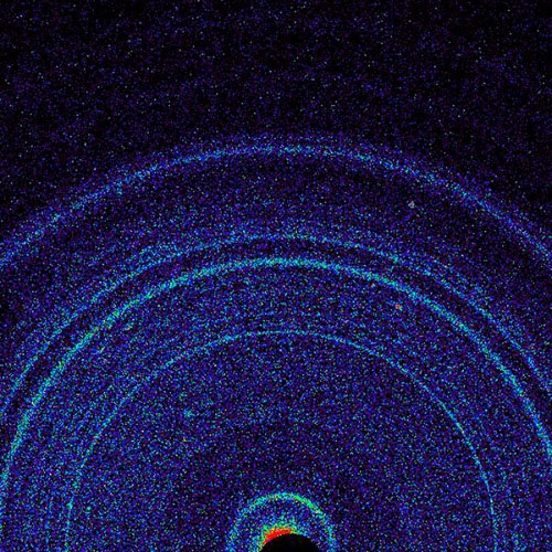 |
| An x-ray diffraction pattern obtained from a sample of Martian soil. X-ray diffraction is a powerful tool for determining the structure of crystalline samples. (Image: NASA/JPL-CALTECH/AMES/SCIENCE PHOTO LIBRARY) |
| The past decade has seen the development of massive x-ray laser facilities known as x-ray free-electron lasers (XFELs) that can deliver extremely intense, ultrashort pulses of x-rays. The pulses are so intense that they destroy samples. But because they are so short, the diffraction pattern can be recorded before a sample blows apart. |
| Recent advances in XFEL technology have allowed a more than 1020-fold increase in the intensity of x-ray beams at the sample surface compared with x-ray tubes used for medical applications, allowing diffraction patterns to be obtained from smaller crystals than was previously possible. But some simulations have predicted that the intensity of diffraction patterns will fall off at such high irradiation intensities. |
| Now, Ichiro Inoue of the RIKEN SPring-8 Center and co-workers have shown experimentally that this is indeed the case (Physical Review Letters, "Femtosecond reduction of atomic scattering factors triggered by intense X-ray pulse"). The team used SACLA in Hyogo prefecture, Japan—one of about five XFELs globally—to measure the diffraction patterns of thin slices of silicon over a range of x-ray intensities. They observed a sharp reduction of nearly 50% above a certain intensity. |
| The reduction was so great that the team picked up on it while performing the experiments, even before analyzing the data. “We could clearly see the drop in diffraction intensity while increasing the x-ray intensity,” recalls Inoue. “That was really surprising.” |
| Simulations performed by the team indicated that above a certain x-ray intensity, the first x-rays to hit the silicon sample knock out electrons lying close to the nuclei of the silicon atoms. As a result of this, subsequent x-rays are scattered much less than usual. |
| This effect will mean that x-ray diffraction patterns obtained at such high intensities will need to be analyzed differently from those obtained at lower intensities. |
| But the effect could be harnessed to create even shorter x-ray pulses. Since the intensity drops off in the latter half of the pulse, silicon could be used as a filter that trims pulses. “We now want to try to demonstrate power shortening based on this phenomenon,” says Inoue. |
| Source: RIKEN (Note: Content may be edited for style and length) |

