| Sep 04, 2019 |
Biophysics: Stretching proteins with magnetic tweezers
|
|
(Nanowerk News) As the central mediators of cell function in biological organisms, proteins are involved in the execution of virtually all cellular processes. They provide the internal scaffolding that gives cells their form, and enable cells to dynamically alter their morphology. They transport substrates back and forth across membranes, and they catalyze most of the chemical reactions that take place in cells. In the course of these tasks many proteins are subjected to external forces.
|
|
Indeed, some “mechanosensitive” proteins effectively measure the strength of the forces acting upon them and are activated when the imposed force exceeds a given threshold value. Von Willebrand Factor (VWF), which initiates the formation of blood clots, is an important representative of this class.
|
|
The mechanical forces required to activate proteins like VWF are often so small that their magnitude could not be determined using existing methods.
|
|
Now, a team of scientists led by LMU physicists Martin Benoit and Professor Jan Lipfert has developed a much more sensitive procedure. Their ‘magnetic tweezers’ can quantify forces that are 100 times smaller than the commonly used alternative method currently available.
|
|
As Lipfert and colleagues report in the journal PNAS ("Multiplexed protein force spectroscopy reveals equilibrium protein folding dynamics and the low-force response of von Willebrand factor"), they have employed the technique to observe the unfolding of the VWF protein under the influence of low mechanical forces.
|
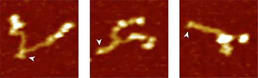 |
| Von Willebrand Factor protein: fully open (left), partially closed and fully closed (right). (Image: Lipfert Group)
|
|
A powerful approach to study mechanoregulation is so-called protein force spectroscopy. This involves tugging on an individual protein molecule and observing how an applied force alters its three-dimensional structure.
|
|
Up to now, the method of choice for pulling has been an atomic force microscope, which works best in the range of 100 piconewton (pN).
|
|
“However, many molecular processes are activated by forces that are much weaker than that,” says Lipfert. “So for measurements at the level of single molecules, we need more sensitive instrumentation – there’s little point in using a bathroom scale to weigh out the ingredients of a cake.”
|
|
The researchers developed a method in which the proteins are attached at one end to a glass surface and carry a tag at the other end that binds to tiny magnetic beads and the assembly is then subjected to an externa magnetic field. Extension of the protein induced by the field results in the vertical displacement of each bead, which can be detected by microscopy.
|
|
“This sort of set-up is referred to as magnetic tweezers,” Lipfert explains. “It has the great advantage that it allows us to apply and resolve very weak forces – significantly less than 1 piconewton – to the protein of interest. In addition, magnetic tweezers enable very stable measurements over long periods of time – up to one week!”
|
|
To test the new method, the LMU group used VWF as their target protein. In the bloodstream, VWF circulates as a multimer of dimers that are made of two identical subunits. Under normal conditions of blood flow, it has a relatively compact globular form.
|
|
However, any increase in the shear forces in the bloodstream owing to injury of the vasculature causes vWF to unfold. This exposes binding sites for receptors on blood platelets. Binding of VWF to platelets in turn triggers a reaction cascade that leads to clotting, which seals the wound.
|
|
“The cascade is induced by the action on the molecule of mechanical forces acting that are much weaker than those that have been measured up to now,” says Lipfert.
|
|
Analysis of the unzipping of VWF dimers with magnetic tweezers showed that the so-called VWF stem opens up under an applied force of less than 1 pN, when the subunits of the dimer are pulled apart like the two halves of a zipper.
|
|
“We assume that this pattern of behavior, which we were able to observe for the first time, represents the first step in blood coagulation,” says Lipfert. “Our approach provides a detailed picture of the forces and the changes in extension involved in unfolding the protein. We are confident that future application of the method will contribute to a better understanding of the mode of action of VWF and of the role of clinically relevant mutations.”
|

