| Aug 31, 2017 |
'Seeing' robot learns tricky technique for studying brain cells in mammals
|
|
(Nanowerk News) Whole-cell patch clamp electrophysiology, or whole-cell recording (WCR), is the gold-standard technique for studying the behaviour of brain cells called neurons under different brain states such as stress or learning.
|
|
The procedure has been used in mammals since it was developed in the 1970s. It helps scientists to understand brain function and brain disorders such as Alzheimer’s. They do this by looking at the electrical activity of individual neurons in a live mammal brain to build a bigger picture of its function as a whole organ. This information is used to understand the role of electrical function in human brain disorders.
|
|
However, WCR is notoriously challenging to perform because of the small scale of the equipment and the microscopic nature of the cells involved. It also requires very precise movements to find neurons and then record their electrical currents accurately. Therefore only a small number of laboratories worldwide specialise in the technique.
|
|
Now, for the first time, a team of scientists led by Professor Simon Schultz and Dr Luca Annecchino at Imperial College London has developed a robot and computer programme that can guide tiny measuring devices called micropipettes to specific neurons in the brains of live mice and record electrical currents, all without human intervention (Neuron, "Robotic Automation of In Vivo Two-Photon Targeted Whole-Cell Patch-Clamp Electrophysiology"). This is the first reported fully automated platform to do this.
|
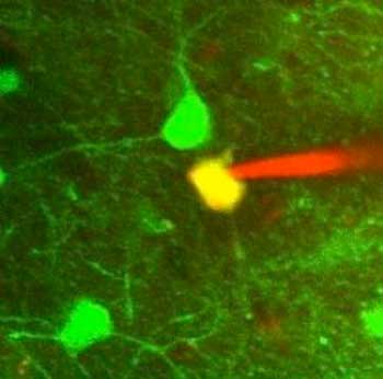 |
| A ‘patched’ neuron: Green shows the fluorescent protein used to guide the robot. Red shows the fluorescent dye inside the pipette. Yellow shows the patched cell. (Image: Imperial College London)
|
|
Senior author Professor Schultz said: “To understand the brain as a whole organ, we need to know how neurons work and communicate with one another. Neurons in themselves are complex structures that use electrical and molecular signals to send information to neighbouring neurons, and the brain as a whole structure. Neurons also act differently depending on whether they are healthy or not fully functioning due to certain brain disorders. The WCR technique is a way to eavesdrop on these cells and how they communicate with their neighbours.
|
|
“However, structures that cannot be seen with the naked human eye require very precise and accurate ways to measure them. We have managed to do so successfully so far, but now we have taught robots to ‘see’ the neuron and perform the procedure even better. This means WCR can now potentially be performed on a much larger scale, which could speed up our learning about the brain and its disorders.”
|
|
The conventional method for carrying out WCR involves scientists tagging a specific neuron with fluorescent protein or dye. They achieve this by guiding a robotic arm to the neuron. This is done by sending electrical pulses into the brain via a pipette filled with electrically conductive fluid. The pulses diffuse into the brain until the micropipette nears a neuron, which creates a block in electrical signal that tells the human or robot operator when to stop moving the micropipette.
|
|
At this point the micropipette clamps onto the outside of the cell, penetrating the membrane using a pulse of suction pressure. It then conducts any electrical signals from the neuron up through the micropipette, and into a computer via the conductive fluid.
|
|
The new method reported in the study by Professor Schultz, from Imperial’s Department of Bioengineering, and his colleagues demonstrates how a robot can do this automatically, without any human input.
|
|
The team compared their technique with the conventional approach and found that the robot was faster and more accurate than its human counterparts. The findings are published in the journal Neuron.
|
|
The automation might mean the technique can be performed much more widely around the world, and even in labs with no expertise in the technique.
|
|
Lead author Dr Annecchino said: “Although the procedure has existed for a number of years, we humans still find it very difficult to perform. However, it is so valuable in teaching us about the mammalian brain, that it is the ideal candidate for robotic automation. We plan to commercialise the programme so that research all over the world can benefit.”
|
|
Next, the researchers will study how brain circuits are disturbed by the amyloid plaques seen in Alzheimer's disease. Dr Annecchino added: “Ultimately, the problems in Alzheimer's result from changes in the information processing capability of networks of individual brain cells. This is exactly what we can monitor with the technique.”
|

