Micro heart muscle created from stem cells (w/video)
(Nanowerk News) Scientists at the Gladstone Institutes have invented a new way to create three-dimensional human heart tissue from stem cells ("Miniaturized iPS-Cell-Derived Cardiac Muscles for Physiologically Relevant Drug Response Analyses"). The tissue can be used to model disease and test drugs, and it opens the door for a precision medicine approach to treating heart disease. Although there are existing techniques to make three-dimensional tissues from heart cells, the new method dramatically reduces the number of cells needed, making it an easier, cheaper, and more efficient system.
"We have bioengineered micro-scale heart tissues with a method that can easily be reproduced, which will enable scientists in stem cell biology and the drug industry to study heart cells in their proper context," said first author Nathaniel Huebsch, PhD, a postdoctoral fellow in the Conklin lab at Gladstone. "In turn, this will enhance our ability to discover treatments for heart disease."
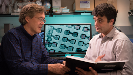
Bruce Conklin and Nathaniel Huebsch have invented a new way to create micro heart muscle from stem cells using a unique dog bone dish. The three-dimensional tissue is ideal for disease modeling and drug testing.
Creating heart cells from induced pluripotent stem cells (iPSCs) that are derived from a patient's skin cells enables scientists to study and test drugs on that patient's specific disease. However, cells made from iPSCs are relatively immature, resembling heart cells in an embryo more than cells in an adult. As such, these cells are inadequate for drug testing because they do not properly predict how a drug will affect adult heart cells. Additionally, heart cells created from iPSCs are challenging to make and work with, so creating large quantities can be difficult. Therefore, the fewer cells needed, the better.
The micro heart muscle addresses both of these concerns. Forcing the cells to organize and stretch into three-dimensional tissue helps spur development and coaxes them into resembling more mature cells that can better predict how a drug will affect adult heart cells. Also, the new method--published in the journal Scientific Reports--requires a thousand-fold fewer cells to grow the tissue than other tissue engineering techniques. Using fewer cells allows the scientists to do many more experiments with the same amount of resources.
3D micro heart muscle created from induced pluripotent stem cells beat in sync.
Working in collaboration with Kevin Healy, PhD, at the University of California, Berkeley, the researchers first generated heart muscle cells and connective tissue cells from iPSCs. They then combined these cells in a special dish shaped like a tiny dog bone. This unique shape encouraged the cells to self-organize into elongated muscle fibers. Within a couple of days, the micro tissues resembled heart muscle both structurally and functionally. For example, when the researchers tested how the tissue responded to certain drugs that impair fetal heart cells but not adult heart cells, the micro heart muscle performed more like adult heart tissue.
"The beauty of this technique is that it is very easy and robust, but it still allows you to create three-dimensional miniature tissues that function like normal tissues," said senior author Bruce Conklin, MD, a senior investigator at Gladstone. "Our research shows that you can create these complex tissues with a simple template that exploits the inherent properties of these cells to self-organize. We think that the micro heart muscle will provide a superior resource for conducting research and developing therapies for heart disease."