Magnetic Force Microscopy (MFM): Probing Magnetic Properties at the Nanoscale
What is Magnetic Force Microscopy (MFM)?
Magnetic Force Microscopy (MFM) is a scanning probe microscopy technique that allows the imaging and characterization of magnetic properties of materials at the nanoscale. MFM is based on the detection of magnetic interactions between a magnetized probe tip and the magnetic fields emanating from a sample surface. This technique provides high-resolution images of magnetic domain structures, magnetic nanoparticles, and other magnetic features with spatial resolutions down to a few tens of nanometers.
Principle of Operation
MFM operates by measuring the magnetic forces between a magnetized probe tip and the magnetic fields emanating from a sample surface. The probe tip is typically coated with a thin magnetic layer, such as cobalt-chromium alloy, and is mounted on a flexible cantilever. As the tip scans over the sample surface, the magnetic interactions cause the cantilever to deflect, which is detected by a laser beam deflection system.
The MFM measurement is usually performed in two passes:
- Topography Pass: In the first pass, the probe tip scans the sample surface in tapping mode or non-contact mode to obtain the topographic information of the surface. The tip-sample distance is maintained at a few nanometers to avoid direct contact with the surface.
- Magnetic Interaction Pass: In the second pass, the probe tip is lifted to a certain height above the sample surface, typically a few tens of nanometers, and scans the same line following the topographic profile obtained in the first pass. At this height, the long-range magnetic forces dominate over the short-range van der Waals forces, allowing the detection of magnetic interactions.
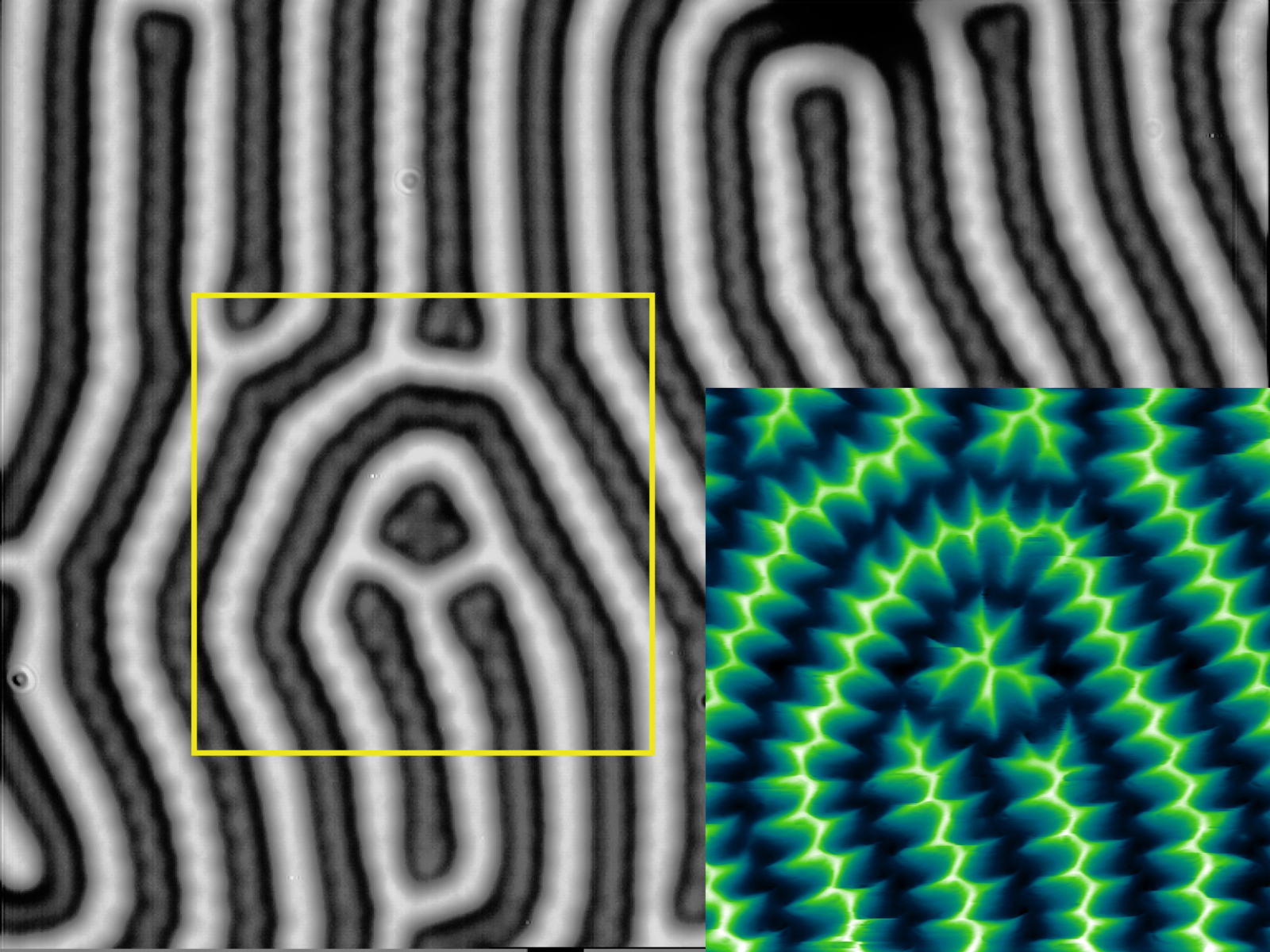
The magnetic forces cause the cantilever to deflect, and the deflection is recorded as a function of the lateral position, generating a magnetic force image of the sample surface. The magnetic contrast in the image arises from the variations in the magnetic properties of the sample, such as magnetic domains, domain walls, and magnetic nanoparticles.
Applications of Magnetic Force Microscopy
MFM has found numerous applications in the field of nanomagnetism and magnetic materials research:
Magnetic Data Storage
MFM is extensively used in the characterization of magnetic recording media, such as hard disk drives and magnetic tapes. It allows the visualization of magnetic bit patterns, domain structures, and the effects of magnetic interactions on the recording performance. MFM imaging helps in optimizing the design and fabrication of high-density magnetic storage devices.
Magnetic Nanoparticles and Nanostructures
MFM is a valuable tool for studying the magnetic properties of individual magnetic nanoparticles and nanostructures. It enables the imaging of magnetic domain configurations, magnetization reversal processes, and the effects of size, shape, and composition on the magnetic behavior of nanoparticles. This information is crucial for the development of magnetic nanomaterials for applications in biomedicine, catalysis, and data storage.
Spintronics and Magnetic Sensors
MFM plays a significant role in the development of spintronic devices and magnetic sensors. It allows the characterization of magnetic thin films, multilayers, and nanostructures used in spintronic applications, such as magnetic random access memories (MRAM) and magnetic field sensors. MFM imaging provides insights into the magnetic switching behavior, domain wall dynamics, and the effects of interfacial coupling in these devices.
Advances and Future Perspectives
Recent advances in MFM have focused on improving the spatial resolution, sensitivity, and functionality of the technique. One approach is the development of high-resolution MFM probes with sharp magnetic tips, such as carbon nanotubes or magnetic nanowires, which enable imaging with resolutions down to a few nanometers. Another area of research is the integration of MFM with other scanning probe techniques, such as Kelvin probe force microscopy and scanning tunneling microscopy, to obtain complementary information about the electronic and magnetic properties of materials.
Future perspectives in MFM include the development of advanced imaging modes, such as 3D magnetic imaging, dynamic MFM, and in-situ MFM under external stimuli (e.g., magnetic fields, temperature, and electrical currents). These advancements will enable the study of complex magnetic phenomena and the exploration of novel magnetic materials and devices.
Further Reading
Magnetochemistry, Magnetic Force Microscopy in Physics and Biomedical Applications
Journal of Applied Physics, Frontiers of magnetic force microscopy
