| Nov 29, 2022 |
These nano-magnets that will restore damaged nerve cells
(Nanowerk News) Neurons are the fundamental units of the brain and nervous system, the cells responsible for receiving sensory input from the external world, for sending motor commands to our muscles, and for transforming and relaying the electrical signals at every step in between. Neurons, also called nerve cells, are composed of three main parts: the cell body, the dendrites and the axon -- a long, thin extension that is responsible for communicating with other cells.
|
|
When neurons are damaged by degenerative disease or injury, they have little, if any, ability to heal on their own. Restoring neural networks and their normal function is therefore a significant challenge in the field of tissue engineering.
|
|
Prof. Orit Shefi and doctoral student Reut Plen from the Kofkin Faculty of Engineering at Bar-Ilan University have developed a novel technique to overcome this challenge using nanotechnology and magnetic manipulations, one of the most innovative approaches to creating neural networks.
|
|
Their research was recently published in the journal Advanced Functional Materials ("Bioengineering 3D Neural Networks Using Magnetic Manipulations").
|
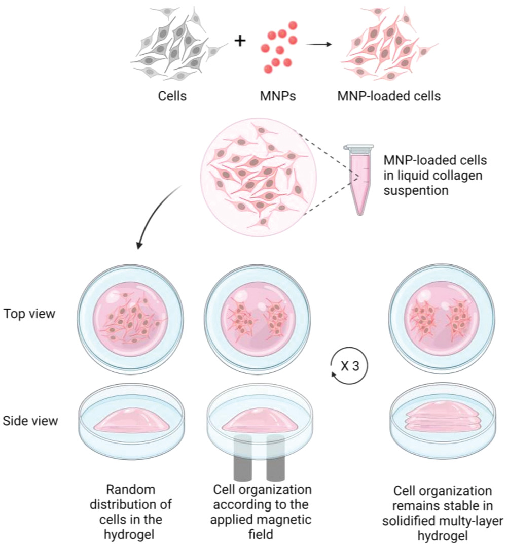 |
| Schematic workflow illustration. Iron oxide nanoparticles are incorporated into cells. MNP-loaded cells are inserted into collagen type-I soft gel and subjected to pre-designed magnetic fields, thus inducing a collective movement toward the magnetic “hot spots” prior to gel solidification. The process is repeated to create a second and third layer of controlled MNP-loaded cells. The spatial organization of the cells is maintained after removing the external magnetic field in the solidified 3D gel. Cells later differentiate and develop neurites, thus creating pre-designed, functional 3D neural networks within the collagen scaffold. (Image: Advanced Functional Materials)
|
|
To create neural networks, the researchers injected magnetic iron oxide nanoparticles into neural progenitor cells, thus turning the cells into independent magnetic units. Next, they exposed the progenitor cells, known to develop into neurons, to a number of pre-adjusted magnetic fields and remotely directed their movement within a three-dimensional and multi-layered collagen substrate that mimics the natural characteristics of body tissue. Through these magnetic manipulations, they created three-dimensional “mini-brains” - functional and multi-layered neural networks that mimic elements found in the brain of mammals.
|
|
After the collagen solution solidified into a gel, the cells remained in place according to the remotely applied magnetic fields. Within a few days, the cells developed into mature neurons, formed extensions and connections, demonstrated electrical activity and thrived in the collagen gel for at least 21 days.
|
|
"This method paves the way for the creation of 3D cell architecture on a customized scale for use in bioengineering, therapeutic and research applications, both inside and outside the body," says PhD student Reut Plen. "Since the 3D neural networks we created simulate innate properties of human brain tissues, they can be used as experimental “mini-brains” and serve as a model for the study of medicinal drugs, for investigating communication between tissues, and as a way to build artificial networks for interfaces between engineering and biological components.
|
|
In addition, the model suggests an interesting prospect for injecting such a gel, which contains cells, in its liquid state, introducing it into the nervous system and organizing the cells into the correct structure with the assistance of magnetic forces. The advantage of using this method is that magnetic fields can affect cells located deep inside the body in a non-invasive manner," adds Plen.
|
|
Inserting magnetic particles into cells, and into nerve cells in particular, raises questions regarding the safety of future medical applications. "The issue of safety is important and we've devoted much thought and research into it," Prof. Orit Shefi points out. "In the first step, we tested the effect of different particles on cell health in culture. In addition, we coated the magnetic particles with a biocompatible protein. The coating creates a buffer between the magnetic element and the cell and encourages penetration of the nanoparticles. Importantly, iron, the building block of the nanoparticle, exists naturally in the body so it isn't a foreign substance. Additionally, the same gel with magnetic particles has been tested in our laboratory and found safe to use in animal models."
|
|
The US Food and Drug Administration has already approved the use of magnetic nanoparticles for diagnostic and imaging purposes and in cases of severe injury. The steps taken by the Bar-Ilan research group create an opportunity to advance the technology for future clinical use. “This is only the beginning,” say Shefi and Plen. “Our novel method of creating ‘mini-brains’ opens the door to finding solutions for various neurological impairments which will hopefully improve the quality of life of numerous patients.
|

