| Apr 26, 2018 |
Noninvasive brain tumor biopsy on the horizon
|
|
(Nanowerk News) Taking a biopsy of a brain tumor is a complicated and invasive surgical process, but a team of researchers at Washington University in St. Louis is developing a way that allows them to detect tumor biomarkers through a simple blood test.
|
|
Hong Chen, a biomedical engineer, and Eric C. Leuthardt, MD, a neurosurgeon, led a team of engineers, physicians and researchers who have developed a groundbreaking, proof-of-concept technique that allows biomarkers from a brain tumor to pass through the tough blood-brain barrier into a patient's blood using noninvasive focused ultrasound and some tiny bubbles, potentially eliminating the need for a surgical biopsy.
|
|
Chen, assistant professor of biomedical engineering in the School of Engineering & Applied Science and of radiation oncology in the School of Medicine, said while researchers have already learned how to get a drug through the blood-brain barrier into the brain via the bloodstream, no one -- until now -- has found a way to release tumor-specific biomarkers -- in this case, messenger RNA (mRNA)-- from the brain into the blood.
|
|
"I see a clear path for the clinical translation of this technique," said Chen, an expert in ultrasound technology. "Blood-based liquid biopsies have been used in other cancers, but not in the brain. Our proposed technique may make it possible to perform a blood test for brain cancer patients."
|
|
The blood test would reveal the amount of mRNA in the blood, which gives physicians specific information about the tumor that can help with diagnosis and treatment options.
|
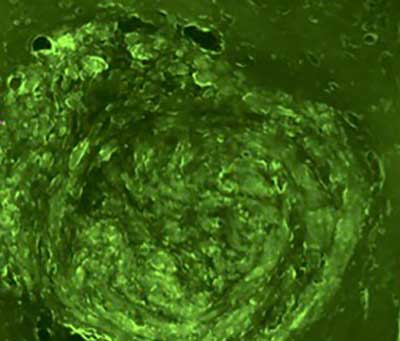 |
| This image shows a brain tumor in a mouse that has been treated with green fluorescent protein-transduced glioblastoma cells. (Image: Washington University in St. Louis)
|
|
Results of the study, which blends imaging, mechanobiology, genomics, immunology, bioinformatics, oncology, radiology and neurosurgery, are published in Scientific Reports ("Focused Ultrasound-enabled Brain Tumor Liquid Biopsy").
|
|
Chen; Leuthardt, professor of neurological surgery in the School of Medicine; and researchers from the schools of Engineering and of Medicine, tested their theory in a mouse model using two different types of the deadly glioblastoma brain tumor. They targeted the tumor using focused ultrasound, a technique that uses ultrasonic energy to target tissue deep in the body without incisions or radiation. Similar to a magnifying glass that can focus sunlight to a tiny point, focused ultrasound concentrates ultrasound energy to a tiny point deep into the brain.
|
|
Once they had the target -- in this case, the brain tumor -- researchers then injected microbubbles that travel through the blood similar to red blood cells. When the microbubbles reached the target, they popped, causing tiny ruptures of the blood-brain barrier that allows the biomarkers from the brain tumor to pass through the barrier and release into the bloodstream. A blood sample can determine the biomarkers in the tumor.
|
|
This technique could lead to personalized medicine.
|
|
"In many ways this has been a holy grail for brain tumor therapy," Leuthardt said. "Having the ability to monitor the changing molecular events of the tumor in an ongoing way allows us to not only better diagnose a tumor in the brain, but to follow its response to different types of treatment."
|
|
"Once the blood-brain barrier is open, physicians can deliver drugs to the brain tumor," Chen said. "Physicians can also collect the blood and detect the expression level of biomarkers in the patient. It enables them to perform molecular characterizations of the brain tumor from a blood draw and guide the choice of treatment for individual patients."
|
|
In addition, Gavin Dunn, MD, assistant professor of neurosurgery, a co-author and leader in cancer immunobiology, plans to use the technique with immunotherapy, which offers precision treatment that targets specific biomarkers in the brain.
|
|
"This noninvasive focused ultrasound-enabled liquid biopsy technique can be useful for long-term monitoring of brain cancer treatment response, where repeated surgical tissue biopsies may not be feasible," Chen said. "Meanwhile, variations within tumors pose a significant challenge to cancer biomarker research. Focused ultrasound can precisely target different locations of the tumor, thereby causing biomarkers to be released in a spatially-localized manner and allow us to better understand the spatial variations of the tumor and develop better treatment." |
|
The team continues to work to refine the process. The future will require integration with advanced genomic sequencing and bioinformatics to enable even more refined diagnostics. These efforts are being led by co-authors, Allegra Petti, assistant professor of medicine, and Xiaowei Wang, associate professor of radiation oncology.
|
|
"Our ongoing work is to optimize the technique and evaluate its sensitivity and safety," Chen said.
|

