| Jul 16, 2018 |
New tools to characterise physical properties of biofilms
|
|
(Nanowerk News) NUS scientists, together with researchers from Nanyang Technological University (NTU) and Imperial College London (ICL), have developed non-invasive biophysical techniques to quantify oxygen concentration and micromechanical properties in bacterial biofilms and understand their real-time responses to environmental changes.
|
|
Communities of bacteria, fungi, protozoa or algae that adhere to each other or surfaces are known as biofilms. The biofilms are characterised by a variety of properties not typically found in isolated free-living organisms. One of the emergent properties of biofilms relevant in the context of human health is their increased tolerance to disinfectants and antibiotics.
|
|
While some biofilms are beneficial (e.g. those involved in wastewater treatment and bioremediation), many others are harmful (e.g. those involved in infections and corrosion). As with any living organism, biofilms continually adapt and respond to a variety of environmental stresses, such as changes in nutrient or oxygen availability.
|
|
Oxygen plays an essential role in the generation of energy for cell maintenance and growth. Quantifying the amount of oxygen is necessary to study its effects during the various stages of biofilm growth. Current tools for measuring oxygen levels in biofilms either consume oxygen themselves (leading to less accurate results) or can only obtain accurate measurements from the surface but not within the biofilms.
|
|
To overcome these limitations, Prof Thorsten WOHLAND from the Departments of Biological Sciences and Chemistry, NUS together with Prof Yehuda COHEN and Prof Scott RICE from NTU have adapted a non-invasive technique called Transient State (TRAST) imaging and applied it to quantify oxygen levels in bacterial biofilms.
|
|
This led to the identification of oxygen deficient zones within the microscopic colonies of P. aeruginosa.
|
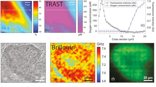 |
| Figures (a) and (b) show the fluorescence and oxygen concentration map of a bacteria microcolony respectively. Figure (c) shows the line profile of fluorescence (circles) and oxygen concentration (triangles) along the dotted line in (a) and (b). Figures (d), (e) and (f) are the wide field microscopic, Brillouin and fluorescence images respectively of a bacteria microcolony showing differences in frequency shift, which can be used to classify the state of the biofilm. (click on image to enlarge)
|
|
TRAST is a luminescence based imaging technique. It is based on the fact that certain fluorophores (a type of fluorescent chemical compound) occupy two different states, one which emits fluorescence and the other a non-fluorescent dark state. The fraction of fluorophores in the dark state depends on how often the fluorophores are excited and whether they are given enough time to come back from the dark states to the fluorescent states.
|
|
By changing the illumination scheme in defined ways, the amount of fluorophores across the biofilm (in the dark state) can be measured easily. The measurements depend only on the fraction of fluorophores in the dark state, meaning that the accuracy is not affected even if certain regions in the biofilm have higher fluorophore concentration. Since oxygen suppresses the occupation of dark states, and thus lowers the fluorophores that reside in the dark state, TRAST can be used to quantify oxygen concentrations.
|
|
This tool has potential implications in microbiology to differentiate oxygen-rich from oxygen-deficient zones, which are typically occupied by aerobic and anaerobic bacteria respectively in a multispecies biofilm. This differentiation is important in diagnostics because this will aid in the identification of the type of bacteria in the infection site.
|
|
The same research team in collaboration with Prof Peter T?R?K from ICL has also developed a technique using Brillouin microscopy to probe the mechanical properties of biofilms at the micrometre scale level. Brillouin microscopy enables the quantification of compressibility by measuring the shift in the frequency of incident light upon interaction with the biofilm. The compressibility of a material is the amount of stress needed to cause a change in the volume of a material.
|
|
The compressibility of a material can be interpreted in terms of the stiffness of the material. Materials that exhibit larger frequency shifts are stiffer than those with smaller frequency shifts. This technique, which is ?label free? (i.e. does not use any foreign molecule(s)), can potentially be used to understand the micromechanical properties of complex biofilms.
|
|
Prof Wohland said, ?Biofilms can have destructive effects, for example in wound infection or in the degradation of materials. However, they can also be harnessed for the production of biological materials or other processes. Both applications need a good understanding of the physical and physiological properties of biofilms. Therefore, new tools, as our team has developed, are needed to better characterise biofilms in their natural surroundings.?
|
Reference
|
|
Karampatzakis A; ; Kandaswamy K; Rice SA; Cohen Y; Wohland T*, "Measurement of oxygen concentrations in bacterial biofilms using transient state monitoring by single plane illumination microscopy" BIOMEDICAL PHYSICS & ENGINEERING EXPRESS Volume: 3 Issue: 3 DOI: 10.1088/2057-1976/aa6db7 Published: 2017.
|
|
Karampatzakis A; Song CZ; Allsopp LP; Filloux A; Rice SA; Cohen Y; Wohland T; Torok P*, "Probing the internal micromechanical properties of Pseudomonas aeruginosa biofilms by Brillouin imaging" NPJ BIOFILMS AND MICROBIOMES Volume: 3 DOI: 10.1038/s41522-017-0028-z Published: 2017.
|
|
Sankaran J; Karampatzakis A; Rice SA; Wohland T*, "Quantitative imaging and spectroscopic technologies for microbiology" FEMS MICROBIOLOGY LETTERS Volume 365 Issue 9 DOI: 10.1093/femsle/fny075 Published: 2018 (Editor?s choice).
|


