| Jun 30, 2021 |
Breakthrough for tracking RNA with fluorescence
|
|
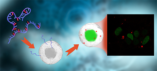
(Nanowerk News) Researchers at Chalmers University of Technology, Sweden, have succeeded in developing a method to label mRNA molecules, and thereby follow, in real time, their path through cells, using a microscope - without affecting their properties or subsequent activity. The breakthrough could be of great importance in facilitating the development of new RNA-based medicines.
|
|
RNA-based therapeutics offer a range of new opportunities to prevent, treat and potentially cure diseases. But currently, the delivery of RNA therapeutics into the cell is inefficient. For new therapeutics to fulfil their potential, the delivery methods need to be optimised.
|
|
Now, a new method, recently presented in the Journal of the American Chemical Society ("Stealth Fluorescence Labeling for Live Microscopy Imaging of mRNA Delivery"), can provide an important piece of the puzzle of overcoming these challenges and take the development a major step forward.
|
|
"Since our method can help solve one of the biggest problems for drug discovery and development, we see that this research can facilitate a paradigm shift from traditional drugs to RNA-based therapeutics," says Marcus Wilhelmsson, Professor at the Department of Chemistry and Chemical Engineering at Chalmers University of Technology, and one of the main authors of the article.
|
 |
| Researchers at Chalmers University of Technology, Sweden, have succeeded in developing a method to label mRNA molecules, and thereby follow, in real time, their path through cells, using a microscope – without affecting their properties or subsequent activity. The breakthrough could be of great importance in facilitating the development of new RNA-based medicines. (Image: Chalmers University of Technology)
|
Making mRNA fluorescent without affecting its natural activity
|
|
The research behind the method has been done in collaboration with chemists and biologists at Chalmers and the biopharmaceuticals company AstraZeneca, through their joint research centre, FoRmulaEx as well as a research group at the Pasteur Institute, Paris.
|
|
The method involves replacing one of the building blocks of RNA with a fluorescent variant, which, apart from that feature, maintains the natural properties of the original base. The fluorescent units have been developed with the help of a special chemistry, and the researchers have shown that it can then be used to produce messenger RNA (mRNA), without affecting the mRNA's ability to be translated into a protein at natural speed.
|
|
This represents a breakthrough which has never before been done successfully. The fluorescence furthermore allows the researchers to follow functional mRNA molecules in real time, seeing how they are taken up into cells with the help of a microscope.
|
|
A challenge when working with mRNA is that the molecules are very large and charged, but at the same time fragile. They cannot get into cells directly and must therefore be packaged. The method that has proven most successful to date uses very small droplets known as lipid nanoparticles to encapsulate the mRNA.
|
|
There is still a great need to develop new and more efficient lipid nanoparticles - something which the Chalmers researchers are also working on. To be able to do that, it is necessary to understand how mRNA is taken up into cells. The ability to monitor, in real time, how the lipid nanoparticles and mRNA are distributed through the cell is therefore an important tool.
|
|
"The great benefit of this method is that we can now easily see where in the cell the delivered mRNA goes, and in which cells the protein is formed, without losing RNA's natural protein-translating ability," says Elin Esbjörner, Associate Professor at the Department for Biology and Biotechnology and the second lead author of the article.
|
Crucial information for optimising drug discovery
|
|
Researchers in this area can use the method to gain greater knowledge of how the uptake process works, thus accelerating and streamlining the new medicines' discovery process. The new method provides more accurate and detailed knowledge than current methods for studying RNA under a microscope.
|
|
"Until now, it has not been possible to measure the natural rate and efficiency with which RNA acts in the cell. This means that you get the wrong answers to the questions you ask when trying to develop a new drug. For example, if you want an answer to what rate a process takes place at, and your method gives you an answer that is a fifth of the correct, drug discovery becomes difficult," explains Marcus Wilhelmsson.
|
On the way to utilisation - directly into IVA's top 100 list
|
|
When the researchers realised what a difference their method could make and how important the new knowledge is for the field, they made their results available as quickly as possible. Recently, the Royal Swedish Academy of Engineering Sciences (IVA) included the project in its annual 100 list and also highlighted it as particularly important for increasing societal resilience to crises. To ensure useful commercialisation of the method, the researchers have submitted a patent application and are planning for a spin-off company, with the support of the business incubator Chalmers Ventures and the Chalmers Innovation Office.
|

