| Oct 10, 2012 |
Nanoscale imaging of strain using X-Ray Bragg projection ptychography |
| (Nanowerk News) The theoretical and experimental framework of a new coherent diffraction strain imaging approach was developed in the Center for Nanoscale Materials' X-Ray Microscopy Group in collaboration with Argonne's Materials Science Division, together with users from IBM. Nanofocused X-ray Bragg projection ptychography creates a tool to efficiently image strain fields with unperturbed boundary conditions in technologically and scientifically relevant energy systems ("Quantitative Nanoscale Imaging of Lattice Distortions in Epitaxial Semiconductor Heterostructures Using Nanofocused X-ray Bragg Projection Ptychography"). |
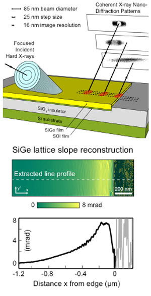 |
| (Top) Focused beam coherent X-ray nanodiffraction patterns collected from a SiGe-on-SOI prototype device edge and (middle and bottom) projected strain field reconstructed by ptychographic methods. |
| This new technique is capable of imaging lattice distortions in thin films nondestructively at spatial resolutions of < 20 nm using coherent nanofocused hard X-rays. This work marks a significant step forward in the development of nondestructive coherent X-ray diffraction imaging techniques for the study of nanoscale lattice features in real materials under real conditions. This study, in which structural subtleties were resolved in a device prototype arising from both intrinsic size effects and extrinsic boundary conditions, paves the way for nondestructive studies of structure in materials at nanometer length scales where prediction, measurement, and control of strain is difficult. |
| The data obtained from the depicted system were used to determine the lattice strain profile in an epitaxial SiGe stressor layer of a silicon prototype device. Measurement of strain from epitaxial lattice mismatches and device processing can test continuum elastic modeling predictions of nanoscale strain distributions. |
| Source: Argonne National Laboratory |

