| Posted: Nov 12, 2008 |
Listening to hot quantum dots adds to nanotherapeutic toolbox |
| (Nanowerk Spotlight) Quantum dots are emerging as an important class of nanoparticles with applications ranging from medicine to energy. These nanocrystals possess size tunable optical and electronic properties resulting from quantum confinement which allow them to be suitable candidates for applications in solar cells and light emitting devices. For instance, quantum dots have been identified as important light harvesting material for building highly efficient solar cells – when exposed to light at certain wavelengths they can generate free electrons and create an electrical current (see: Catching a rainbow - quantum dot nanotechnology brightens the prospects for solar energy). Having a high resistance to photobleaching, quantum dots (QDs) also are attractive materials for optoelectronics and in vivo biosensing (see: Live cell imaging with biodegradable quantum dot nanocomposites). |
| Researchers have now demonstrated that QDs, in addition to being excellent fluorescent probes, can be used as photoacoustic (which combines the advantages of optical absorption contrast with ultrasonic spatial resolution for deep imaging) and photothermal (light absorbed and not lost by emission results in heating, which can be measured) contrast agents and sensitizers, thereby providing an opportunity for multimodal high resolution (300 nm) photoacoustic/photothermal-fluorescent imaging as well as sensitizers in nanotherapeutic applications. |
| "We have demonstrated that part of the absorbed energy – in fact, up to 60% – in quantum dots which can be transformed into heat and then at certain condition into sound can be sufficient for detection of quantum dots using thermal or ultrasound transducers" Dr. Vladimir P. Zharov tells Nanowerk. "These findings may open a new avenue in applying quantum dots as triple contrast agents, which besides fluorescent imaging can be used with so-called photothermal and photoacoustic techniques based on detecting laser-induced heat and acoustic waves, and nano- and microbubbles." |
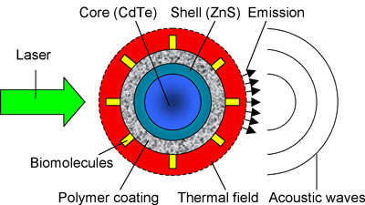 |
| Quantum dots as multimodal contrast agents for integrated fluorescent, photothermal, and photoacoustic detection and imaging. (Image: Dr. Zharov, University of Arkansas for Medical Sciences) |
| Zharov is professor of Biomedical Engineering and Director of the Philips Classic Laser Laboratories at the Winthrop P. Rockefeller Cancer Institute at the University of Arkansas for Medical Sciences. Together with collaborators from the Prokhorov General Physics Institute in Moscow, Russia, the Division of Human Gene Therapy at the University of Alabama, and the Institute of Optics and Biophotonics at Saratov State University, Russia, he has published the team's findings in the October 4, 2008 online edition of Nano Letters ("Quantum Dots as Multimodal Photoacoustic and Photothermal Contrast Agents"). |
| Photothermal and photoacoustic techniques are thought to have great potential for noninvasive in vivo tumor imaging and other pathologies, with higher resolution and in deeper tissue compared to most other optical modalities. It was also believed that these technique are more appropriate for sensing strongly absorbing non-fluorescent objects such as blood vessels or gold nanoparticles. |
| Zharov and his colleagues exploited the fact that, when QDs are irradiated by short laser pulses, multirelaxation processes in the nanoparticles eventually lead to conversion of part of the absorbed energy into heat and subsequent photoacoustic effects. They have shown that, using optimized schematics and advanced technical components, photothermal and photoacoustic methods also have the capability to detect and image quantum dots with a sensitivity and resolution comparable to that of the fluorescence method. |
| "Quantum dots absorb much less light energy compared to strongly absorbing gold nanorods and carbon nanotubes" explains Zharov. "Nevertheless, we observed readable signals at relatively low dot concentrations. Besides a new photoacoustic schematic and using one of the most sensitive ultrasound transducer, we discovered that the structure of a quantum dot can result in an enhancing effect when generating photoacoustic signals. In particular, the protective external layer may play the role of heat accumulator and effective converter of heat into acoustic waves." |
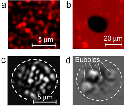 |
| High (a, 60 x) and low (b, 10 x ) resolution fluorescence images of QDs before (a) and after (b) one laser pulse (532 nm, 3.5 J/cm2, 20 µm diameter beam); excitation: 465 nm, emission: 665 nm. Note: the higher concentration of QDs and lower optical resolution in (b) do not allow to distinguish single QDs (just a few large clusters) compared to that of (a). PT images at low (c) and high (d) laser fluence: 0.6 J/cm and 3.5 J/cm2, respectively. PT (c) and fluorescent (a) images were obtained from different parts of the same sample. Dash curve in (d) indicates laser beam diameter. (Image: Dr. Zharov, University of Arkansas for Medical Sciences) |
| The observations made by Zharov's team may significantly extend the traditional application of QDs and these novel dual fluorescent/photoacoustic or fluorescent/photothermal modalities may find many biological application. Zharov also points out that, in contrast to fluorescence, the photoacoustic signals did not show any blinking behavior (i.e. when signal amplitude is not stable over time) and can be detectable even from QDs which do not exhibit readable fluorescence, i.e., they are invisible with fluorescence techniques. |
| Dr. Ekaterina Galanzha, who is a medical co-worker in this study, mentions several possible examples for imaging applications: "In vivo studies that analyze QD pharmokinetics (e.g., clearance and accumulation rates, tissue biodistribution) in blood, the lymphatic system as well as tissues and organs up to 2-3 cm deep that are accessible with photoacoustic techniques; studies using fluorescent/photothermal/photoacoustic imaging for guiding photothermal therapy, including tracking of individual tumor cells, bacteria or viruses labeled with bioconjugated quantum dots; mapping of tumor margins, sentinel lymph nodes and infected sites with integrated imaging modalities, or the use for multimodal molecular targeting and imaging in vivo blood and lymph flow cytometry." |
| Another example is the use of quantum dots as thermal sensitizer or therapeutic agents where the laser-induced high temperature around quantum dots, accompanied by a bubble formation phenomena, can damage surrounding tissue and kill the affected cancer cells. Interestingly, the toxicity concerns that accompany the use of quantum dots can be an actual advantage in such an application because laser-induced removal of the protective external layer around the QD core may provide a synergistic enhancement of the cell-killing (simultaneous thermal, bubble and toxic) effects for deadly infections or tumors. |
| Laser-induced microbubbles around overheated QDs may create asymmetric forces that can move QDs away from the laser beam – the principle used in photoacoustic tweezers that were recently developed by Zharov ("Photoacoustic tweezers with a pulsed laser: theory and experiments"). A relatively high laser energy may inhibit QD fluorescence through a so-called photothermal bleaching mechanism (see the 'black hole' in image 'b' in the above figure), that can be used for laser dosimetry and photosafety standard for quantum dots. |
| Based on this recent and previous work, the scientists have identified several ways for further improving quantum dots' photothermal and photoacoustic contrasts without influence of fluorescent properties, including changing the laser pulse duration or using special coatings with optimized thermal and acoustic parameters. With pulse lasers with different wavelengths – 'laser rainbow' – and a time- resolved detection technique ("In vivo multispectral, multiparameter, photoacoustic lymph flow cytometry with natural cell focusing, label-free detection and multicolor nanoparticle probes"), QDs with different absorption spectra can also be used as photothermal and photoacoustic multicolor contrast agents. |
| "I think that the combination of a time-resolved two-beam technique (pump-beam and probe-beam beam with different time delays between pump and probe laser pulses) with picosecond and especially, femtosecond lasers – so far we have used a nanosecond laser – with photothermal and photoacoustic detection schematics may increase significantly our knowledge of physical effects in quantum dots and light-dot interaction. It may include rapid assessment of quantum yields of QDs, quenching phenomena (i.e., inhibition of fluorescence) in different environments or specific relaxation times in QDs — i.e., time scale of transformation of absorbed energy in fluorescence, heat and acoustic energy" says Zharov. |
 By
Michael
Berger
– Michael is author of four books by the Royal Society of Chemistry:
Nano-Society: Pushing the Boundaries of Technology (2009),
Nanotechnology: The Future is Tiny (2016),
Nanoengineering: The Skills and Tools Making Technology Invisible (2019), and
Waste not! How Nanotechnologies Can Increase Efficiencies Throughout Society (2025)
Copyright ©
Nanowerk LLC
By
Michael
Berger
– Michael is author of four books by the Royal Society of Chemistry:
Nano-Society: Pushing the Boundaries of Technology (2009),
Nanotechnology: The Future is Tiny (2016),
Nanoengineering: The Skills and Tools Making Technology Invisible (2019), and
Waste not! How Nanotechnologies Can Increase Efficiencies Throughout Society (2025)
Copyright ©
Nanowerk LLC
|

Become a Spotlight guest author! Join our large and growing group of guest contributors. Have you just published a scientific paper or have other exciting developments to share with the nanotechnology community? Here is how to publish on nanowerk.com.
