| Posted: Dec 05, 2016 |
DNA + nanoparticles = self-assembled 'diamond'
(Nanowerk News) The remarkable properties that diamonds possess result from their crystalline structure; similar structures in which nanoparticles substitute for carbon atoms could lead to materials with new and undiscovered properties.
|
|
For the first time, scientists realized ordered arrays (superlattices) of nanoparticles, which self-organize in the same way that carbon atoms do within a diamond lattice (Science, "Diamond family of nanoparticle superlattices").
|
|
This new self-organization scheme traps nanoparticles in self-assembled DNA cages that then form large, ordered nanoparticle assemblies.
|
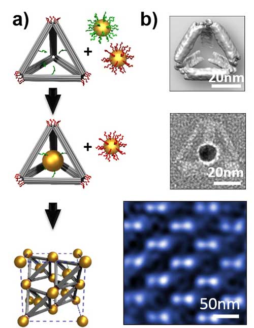 |
| a) Illustration of the approach used to fabricate a diamond superlattice with gold nanoparticles and DNA.b)Experimental results: Top: Cryo-transmission electron microscopy density map of a DNA tetrahedron. Middle: Transmission electron microscopy image of a DNA-caged nanoparticle (scale bars, 20 nm). Bottom: High magnification scanning transmission electron microscopy image of the diamond superlattice along the [110] zone axis (scale bar, 50 nm). (Image: Oleg Gang)
|
|
This novel method of using DNA as linkers of and cages for nanoparticles redefines the rules for connecting nanoparticles and opens exciting prospects for the bottom-up creation of complex nanomaterials with yet undiscovered properties for applications in energy conversion, energy storage, and computing applications, among others.
|
|
For the first time, scientists devised ordered arrays (superlattices) of nanoparticles, which organize in the same way that carbon atoms do within the diamond lattice.
|
|
They achieved these diamond-type superlattices by associating DNA-functionalized gold nanoparticles with “guest” nanoparticles that were encaged by tetrahedral frames made from rigid DNA bundles.
|
|
By varying the size or species of the “guests,” the team produced variants of the diamond lattice, with and without an analog in atomic lattices.
|
|
The team used the Center for Functional Nanomaterials’ Materials Synthesis & Characterization and Electron Microscopy Facilities to produce the superlattices and to characterize their structures using electron microscopy methods (transmission electron microscopy (TEM), scanning TEM, and cryo-TEM).
|
|
The team used the Center for Functional Nanomaterials’ Advanced X-ray Probes Facility at the National Synchrotron Light Source to acquire small-angle x-ray scattering data; they used the Theory and Computation Facility for modeling.
|