Stochastic Optical Reconstruction Microscopy (STORM): Unlocking the Nanoscale World
What is STORM?
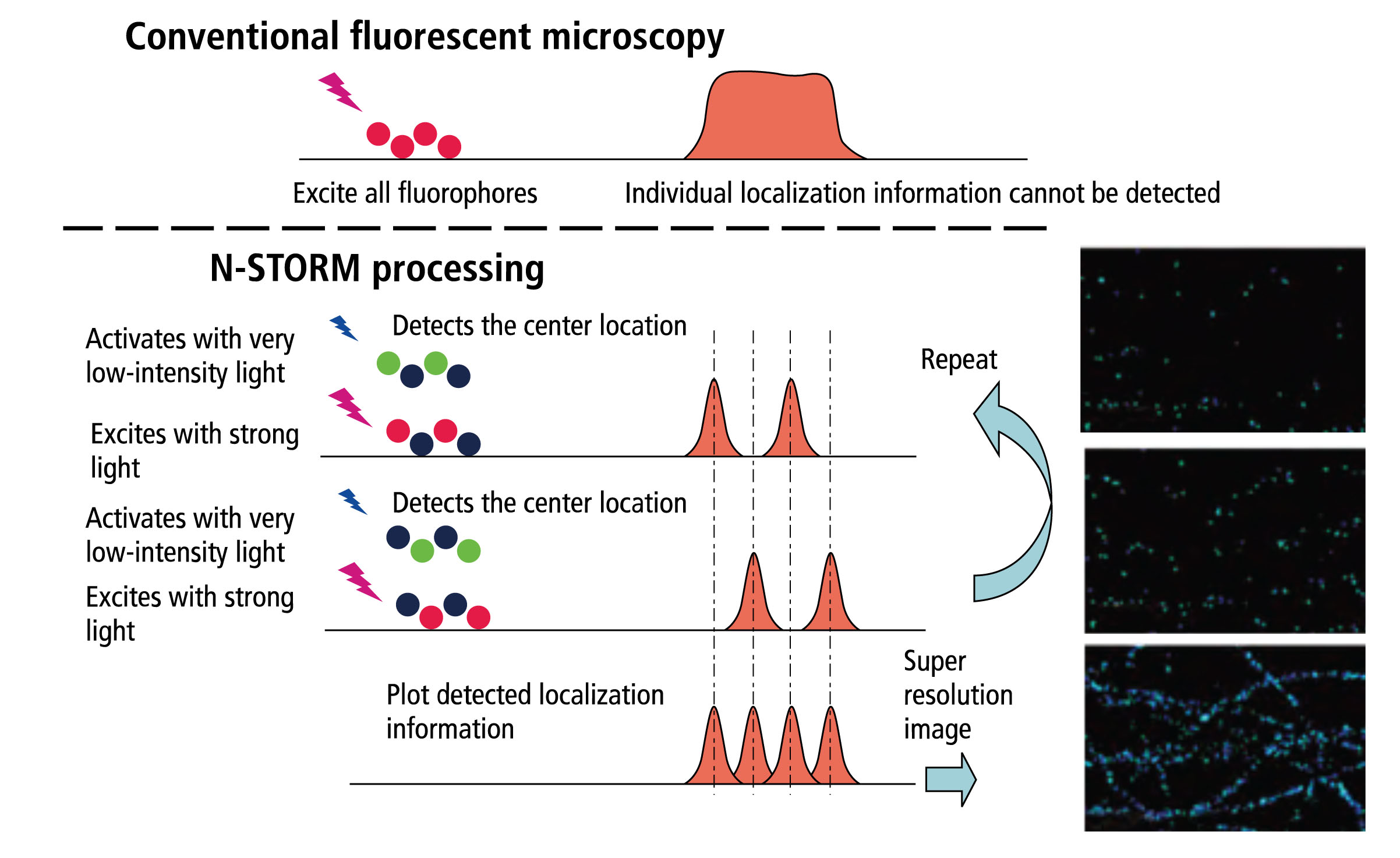
Stochastic Optical Reconstruction Microscopy (STORM) is a super-resolution imaging technique that enables the visualization of biological structures and processes at the nanoscale. It overcomes the diffraction limit of conventional optical microscopy, providing a resolution of approximately 20 nanometers, which is about 10 times better than traditional light microscopy.
Principles of STORM
STORM relies on the stochastic activation and precise localization of individual fluorescent molecules to reconstruct a super-resolution image. The key principles of STORM include:
Photoswitchable Fluorophores
STORM uses special fluorescent probes, called photoswitchable fluorophores, that can be switched between a fluorescent "on" state and a dark "off" state. These fluorophores are typically organic dyes or fluorescent proteins that can be controlled by light of different wavelengths.
Stochastic Activation
During imaging, the photoswitchable fluorophores are randomly activated, such that only a sparse subset of them is in the fluorescent state at any given time. This stochastic activation allows for the precise localization of individual fluorophores without overlap.
Precise Localization
The position of each activated fluorophore is determined with high precision by fitting its emission pattern to a 2D Gaussian function. By repeating the activation and localization process over multiple cycles, a large number of fluorophore positions are obtained.
Image Reconstruction
The final super-resolution image is reconstructed by plotting the positions of all the localized fluorophores. The resulting image reveals the underlying structure with a resolution far beyond the diffraction limit.
Advantages of STORM
STORM offers several advantages over other super-resolution techniques:
- High spatial resolution, typically around 20 nm in the lateral direction and 50 nm in the axial direction.
- Compatibility with a wide range of fluorescent probes, including organic dyes and fluorescent proteins.
- Ability to image multiple targets simultaneously using different fluorophore colors.
- Relatively simple and cost-effective instrumentation compared to other super-resolution methods.
Applications of STORM
STORM has found numerous applications in biological and biomedical research, enabling the study of nanoscale structures and processes in unprecedented detail. Some key applications include:
Cellular Imaging
STORM has been widely used to image subcellular structures, such as cytoskeletal filaments, organelles, and protein complexes. It has provided new insights into the organization and dynamics of these structures at the nanoscale.
Protein Localization and Interaction
STORM allows for the precise localization of proteins within cells and the study of protein-protein interactions. By labeling proteins with different fluorophores, their spatial relationships and co-localization can be determined with high resolution.
Molecular Mechanisms of Disease
STORM has been applied to investigate the molecular basis of various diseases, such as neurodegenerative disorders, cancer, and infectious diseases. It has revealed abnormalities in protein aggregation, cellular structures, and pathogen-host interactions at the nanoscale.
Drug Discovery and Development
STORM can be used to study the effects of drugs on cellular structures and processes at the molecular level. It can provide valuable information for drug target identification, lead optimization, and mechanistic understanding of drug action.
Challenges and Future Perspectives
Despite its powerful capabilities, STORM also faces several challenges. One major limitation is the requirement for specialized fluorescent probes that can undergo photoswitching. The development of new and improved fluorophores with enhanced brightness, photostability, and switching properties is an active area of research.
Another challenge is the need for robust data analysis and image reconstruction algorithms to handle the large amounts of localization data generated by STORM. Advances in computational methods, such as machine learning and artificial intelligence, are expected to streamline and improve the image reconstruction process.
In the future, STORM is likely to be combined with other imaging modalities, such as electron microscopy and cryo-electron tomography, to provide a more comprehensive understanding of biological systems across multiple scales. The integration of STORM with live-cell imaging techniques will also enable the study of dynamic processes in real-time with nanoscale resolution.
Further Reading
Nature Methods, Sub-diffraction-limit imaging by stochastic optical reconstruction microscopy (STORM)
Current Protocols in Cytometry, Stochastic optical reconstruction microscopy (STORM)
Coordination Chemistry Reviews, Organic fluorescent probes for stochastic optical reconstruction microscopy (STORM): Recent highlights and future possibilities
