Super-Resolution Microscopy: Unveiling the Nanoscale World
What is Super-Resolution Microscopy?
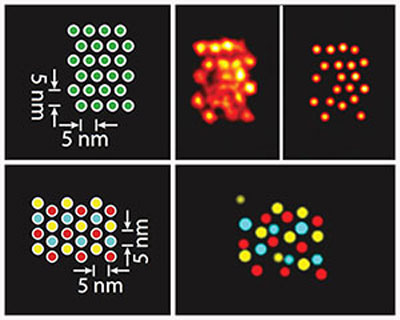
Super-resolution microscopy is a set of advanced optical imaging techniques that overcome the diffraction limit of conventional light microscopy, enabling the visualization of structures and processes at the nanoscale. By employing various strategies to manipulate light and fluorescent probes, super-resolution microscopy allows researchers to observe biological and material systems with unprecedented spatial resolution, often down to a few nanometers.
Breaking the Diffraction Limit
The diffraction limit is a fundamental barrier in conventional light microscopy, restricting the spatial resolution to about half the wavelength of the illuminating light (typically around 200-300 nm). This limit arises from the wave nature of light and the inability to focus light to an infinitely small point. Super-resolution microscopy techniques overcome this limit through various approaches:
- Structured Illumination Microscopy (SIM): Structured Illumination Microscopy uses patterned illumination to encode high-frequency spatial information into observable low-frequency signals. By capturing multiple images with different illumination patterns and computationally reconstructing the high-resolution image, SIM can achieve a twofold improvement in spatial resolution compared to conventional microscopy.
- Stimulated Emission Depletion (STED) Microscopy: Stimulated Emission Depletion Microscopy employs a doughnut-shaped depletion laser to selectively switch off fluorophores around the edges of the excitation spot. By scanning the sample with this reduced excitation volume, STED can achieve spatial resolutions down to 20-30 nm, a tenfold improvement over the diffraction limit.
- Single-Molecule Localization Microscopy (SMLM): Single-Molecule Localization Microscopy techniques, such as Photoactivated Localization Microscopy (PALM) and Stochastic Optical Reconstruction Microscopy (STORM), rely on the sequential activation and precise localization of individual fluorophores. By reconstructing the positions of thousands of single molecules over multiple frames, SMLM can generate super-resolution images with a spatial resolution of 10-20 nm.
Applications of Super-Resolution Microscopy
Super-resolution microscopy has revolutionized our understanding of biological and material systems at the nanoscale. Some key applications include:
Cellular and Molecular Biology
Super-resolution microscopy has enabled the visualization of previously unresolved cellular structures and processes, such as the organization of protein complexes, the dynamics of membrane receptors, and the architecture of the cytoskeleton. By providing nanoscale insights into cellular function, super-resolution microscopy has shed light on fundamental biological questions and disease mechanisms.
Neuroscience
Super-resolution microscopy has been instrumental in studying the complex organization and function of neural circuits. It has allowed researchers to map the nanoscale structure of synapses, trace the connectivity of individual neurons, and investigate the molecular mechanisms underlying synaptic plasticity and neurodegenerative disorders.
Materials Science
Super-resolution microscopy has found applications in the characterization of nanostructured materials, such as nanoparticles, 2D materials, and thin films. By providing nanoscale imaging capabilities, super-resolution techniques have enabled the study of surface morphology, defects, and optical properties of materials with unprecedented detail.
Challenges and Future Perspectives
Despite the remarkable advances in super-resolution microscopy, several challenges remain. One of the main challenges is the trade-off between spatial resolution, temporal resolution, and imaging depth. Achieving high spatial resolution often comes at the cost of slower imaging speeds and limited penetration depth in thick samples. Ongoing research aims to develop faster and more robust super-resolution techniques that can image deeper into tissues and capture dynamic processes in real-time.
Another challenge is the need for specialized fluorescent probes and labeling strategies that are compatible with super-resolution imaging. The development of brighter, more photostable, and switchable fluorophores will expand the applicability of super-resolution microscopy to a wider range of biological systems and research questions.
Future developments in super-resolution microscopy will likely involve the integration of multiple imaging modalities, such as combining super-resolution with other techniques like electron microscopy, atomic force microscopy, or Raman spectroscopy. This multimodal approach will provide a more comprehensive understanding of nanoscale structures and processes, bridging the gap between molecular and cellular scales.
Further Reading
Journal of Cell Biology, A Guide to Super-Resolution Fluorescence Microscopy
Trends in Cell Biology, Super-Resolution Microscopy: From Single Molecules to Supramolecular Assemblies
