Structured Illumination Microscopy (SIM): Enhancing Resolution in Fluorescence Microscopy
What is Structured Illumination Microscopy?
Structured Illumination Microscopy (SIM) is a super-resolution fluorescence microscopy technique that allows for imaging beyond the diffraction limit of light. It works by projecting a known pattern of light (structured illumination) onto the sample, which mixes with the sample's features to create moiré fringes. By capturing multiple images with the illumination pattern shifted to different positions and orientations, a high-resolution image can be computationally reconstructed, effectively doubling the resolution compared to conventional microscopy.
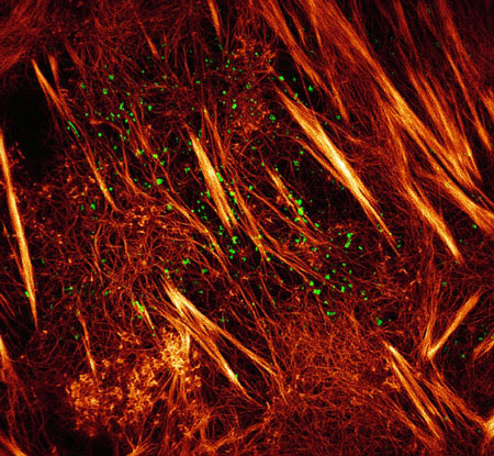
Here is a revised caption with less jargon for the structured illumination microscopy (SIM) image:
Principle of Structured Illumination
The key concept behind SIM is the use of structured illumination to encode high-resolution information into the observed image. By projecting a known pattern (usually a sinusoidal grid) onto the sample, moiré fringes are created due to the interference between the illumination pattern and the sample's features. These moiré fringes contain information about the sample's high-frequency details that are normally lost in conventional microscopy due to the diffraction limit.
To extract the high-resolution information, multiple images are acquired with the illumination pattern shifted to different positions and orientations. Typically, nine images are captured (three phase shifts for each of three pattern orientations). These images are then processed using a specialized algorithm that separates the high-frequency information from the moiré fringes and reconstructs a super-resolution image.
Advantages of SIM
SIM offers several advantages over conventional fluorescence microscopy:
- Resolution Enhancement: SIM can achieve a lateral resolution of approximately 100 nm, which is twice the resolution of conventional microscopy. This allows for the visualization of finer details and structures in biological samples.
- Optical Sectioning: SIM provides optical sectioning capabilities, enabling the acquisition of 3D images with reduced out-of-focus background. This is particularly useful for imaging thick samples or live cells.
- Compatibility with Standard Fluorophores: SIM is compatible with most standard fluorescent labels and dyes used in biological imaging. This makes it easily adaptable to existing sample preparation protocols and allows for multicolor imaging.
- Live Cell Imaging: SIM is relatively fast compared to other super-resolution techniques, making it suitable for live cell imaging. It can capture dynamic processes with a temporal resolution of a few seconds per frame.
Comparison with Other Super-Resolution Techniques
SIM is one of several super-resolution microscopy techniques that have been developed to overcome the diffraction limit. Here's a comparison with two other popular techniques:
Stimulated Emission Depletion (STED) Microscopy
STED microscopy achieves super-resolution by selectively deactivating fluorophores around a focal point using a doughnut-shaped depletion beam. This results in a smaller effective excitation spot, enabling higher resolution. STED can achieve a lateral resolution of 20-50 nm, surpassing SIM. However, STED requires specialized fluorophores and high laser intensities, which can be more challenging for live cell imaging.
Single-Molecule Localization Microscopy (SMLM)
SMLM techniques, such as Photoactivated Localization Microscopy (PALM) and Stochastic Optical Reconstruction Microscopy (STORM), rely on the sequential activation and localization of individual fluorophores to reconstruct a super-resolution image. By precisely determining the positions of individual molecules, SMLM can achieve a lateral resolution of 10-30 nm. However, SMLM requires longer acquisition times and specialized fluorophores, making it less suitable for fast live cell imaging compared to SIM.
In summary, SIM offers a balance between resolution enhancement, speed, and compatibility with standard fluorophores, making it a versatile tool for biological imaging. While STED and SMLM can provide higher resolution, SIM's relatively simple setup and fast imaging capabilities make it an attractive choice for many applications.
Applications of SIM
SIM has been applied to a wide range of biological studies, including:
- Visualizing subcellular structures, such as the cytoskeleton, organelles, and protein complexes
- Studying protein-protein interactions and co-localization at the nanoscale
- Investigating dynamic processes, such as membrane trafficking and cell division
- Imaging 3D structures in thick samples or tissue sections
SIM's ability to provide high-resolution images with optical sectioning has made it a valuable tool for understanding the nanoscale organization and dynamics of biological systems.
Further Reading
Advances in Optics and Photonics, Structured illumination microscopy
Frontiers in Physics, Advances in High-Speed Structured Illumination Microscopy
