Electron Microscopy: Unveiling the Nanoscale World
What is Electron Microscopy?
Electron microscopy is a powerful imaging technique that utilizes a beam of accelerated electrons to visualize and analyze the structure, composition, and properties of materials at the nanoscale. Unlike traditional light microscopes, which are limited by the wavelength of visible light, electron microscopes can achieve much higher magnifications and resolutions, enabling researchers to observe and study objects that are too small to be seen with optical microscopes.
Types of Electron Microscopes
There are two main types of electron microscopes:
Transmission Electron Microscope (TEM)
In a Transmission Electron Microscope, a beam of electrons is transmitted through an ultrathin sample, and the interactions between the electrons and the sample create an image. TEM provides high-resolution images of the internal structure of materials, with magnifications up to several million times. It is widely used for the characterization of nanoparticles, nanostructures, and biological samples.
Scanning Electron Microscope (SEM)
A Scanning Electron Microscope uses a focused beam of electrons to scan the surface of a sample, generating high-resolution images of the sample's topography and composition. The electrons interact with the atoms in the sample, producing various signals that can be detected and used to create an image or analyze the sample's chemical composition. SEM has a lower resolution compared to TEM but provides detailed surface information and has a larger depth of field.
Key Components of Electron Microscopes
Electron microscopes consist of several key components:
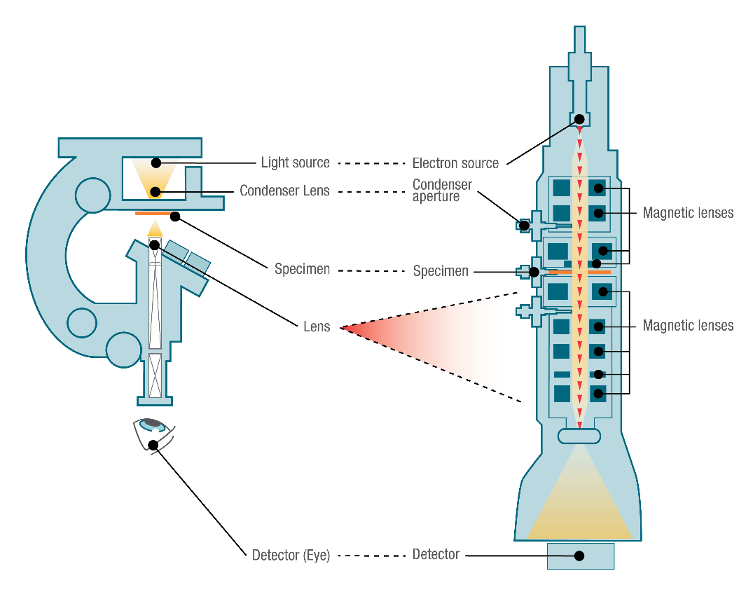
- Electron Gun: The electron gun generates a beam of electrons by thermionic emission or field emission. The electrons are accelerated to high energies, typically ranging from a few keV to several hundred keV.
- Electromagnetic Lenses: Electron microscopes use electromagnetic lenses to focus and manipulate the electron beam. These lenses are analogous to the glass lenses used in light microscopes but use magnetic fields to control the path of the electrons.
- Sample Stage: The sample stage holds the specimen being imaged and allows for precise positioning and manipulation of the sample. In TEM, the sample must be ultrathin (typically less than 100 nm) to allow the electrons to pass through, while SEM can image thicker samples.
- Detectors: Electron microscopes are equipped with various detectors to collect the signals generated by the interaction of the electron beam with the sample. These detectors include secondary electron detectors, backscattered electron detectors, and X-ray detectors, which provide different types of information about the sample.
Applications of Electron Microscopy in Nanotechnology
Electron microscopy plays a crucial role in the field of nanotechnology, enabling researchers to visualize, characterize, and manipulate materials at the nanoscale:
Nanomaterial Characterization
Electron microscopy is extensively used for the characterization of nanomaterials, such as nanoparticles, nanowires, and nanotubes. It provides information about their size, shape, morphology, and crystal structure, which are essential for understanding their properties and behavior.
Nanodevice Fabrication and Analysis
Electron microscopy is indispensable for the fabrication and analysis of nanodevices, such as nanoelectronic circuits, nanophotonic devices, and nanoelectromechanical systems (NEMS). It enables the imaging and characterization of nanoscale features, defects, and interfaces, ensuring the quality and functionality of these devices.
Biological and Biomedical Applications
Electron microscopy is widely used in the biological and biomedical sciences for the study of viruses, bacteria, cells, and tissues at the nanoscale. It provides detailed insights into the structure and organization of biological systems, aiding in the development of targeted drug delivery systems, diagnostic tools, and therapeutic interventions.
Advances and Future Perspectives
Electron microscopy continues to evolve, with ongoing developments in instrumentation, data acquisition, and image processing techniques. Some of the recent advances include aberration-corrected electron microscopy, which enables sub-angstrom resolution imaging, and in situ electron microscopy, which allows for the real-time observation of dynamic processes at the nanoscale.
Future research in electron microscopy will focus on pushing the boundaries of resolution, sensitivity, and speed. The integration of electron microscopy with other analytical techniques, such as spectroscopy and diffraction, will provide a more comprehensive understanding of nanomaterials and nanostructures. Additionally, the development of automated data acquisition and analysis methods will streamline the imaging process and enhance the reproducibility and throughput of electron microscopy experiments.
Further Reading
Nature Reviews Methods Primers, Volume electron microscopy
Clinical Microbiology Reviews, Modern Uses of Electron Microscopy for Detection of Viruses
