Electron Tomography: Unveiling 3D Nanostructures with Atomic Precision
What is Electron Tomography?
Electron tomography is a powerful imaging technique that enables the three-dimensional (3D) visualization and analysis of nanostructures at the atomic scale. By combining advanced electron microscopy with computational reconstruction algorithms, electron tomography provides unprecedented insights into the internal structure, composition, and properties of nanomaterials and biological specimens.
Principle of Electron Tomography
Electron tomography is based on the principle of tomographic reconstruction, which involves acquiring a series of two-dimensional (2D) projection images of a specimen at different tilt angles using an electron microscope. These projection images are then computationally combined to reconstruct a 3D volume of the specimen, revealing its internal structure with nanoscale resolution.
The key steps in electron tomography are:
- Sample Preparation: The specimen is prepared for electron microscopy, typically by embedding it in a resin or by using cryo-electron microscopy techniques to preserve its native structure.
- Data Acquisition: A series of 2D projection images are acquired at different tilt angles using an electron microscope, such as a transmission electron microscope (TEM) or a scanning transmission electron microscope (STEM). The tilt angles typically range from -70° to +70° with increments of 1-2°.
- Alignment and Reconstruction: The acquired projection images are aligned to correct for any specimen drift or mechanical instabilities during data acquisition. Then, computational algorithms, such as weighted back-projection (WBP) or iterative reconstruction methods, are used to reconstruct a 3D volume from the aligned projection images.
- Visualization and Analysis: The reconstructed 3D volume is visualized using advanced software tools, allowing researchers to explore the internal structure of the specimen, measure distances and angles, and perform quantitative analyses.
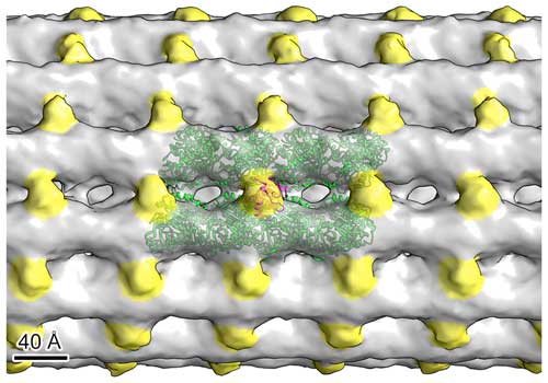
Cryo-Electron Tomography
Cryo-electron tomography (cryo-ET) is a specialized technique that combines electron tomography with cryo-electron microscopy (cryo-EM) to study the 3D structure of biological specimens in their native, hydrated state. Unlike conventional electron tomography, which often requires samples to be fixed, dehydrated, and stained, cryo-ET preserves the specimen's native structure by rapidly freezing it in a thin layer of vitreous ice.
The main advantages of cryo-ET over conventional electron tomography for biological specimens are:
- Preservation of Native Structure: Cryo-ET allows the visualization of biological structures in their near-native state, without the artifacts introduced by chemical fixation, dehydration, or staining.
- Improved Contrast: The rapid freezing of specimens in cryo-ET enhances the inherent contrast of biological structures, eliminating the need for heavy metal staining and enabling the visualization of low-contrast features.
- Reduced Radiation Damage: The cryogenic temperatures used in cryo-ET help to minimize radiation damage to the specimen during imaging, allowing for longer exposure times and higher electron doses.
Cryo-ET has revolutionized the field of structural biology by enabling the 3D visualization of macromolecular complexes, organelles, and even entire cells in their native cellular context. It has provided unprecedented insights into the structure and function of viruses, the nuclear pore complex, the mitochondrial inner membrane, and many other biological systems.
However, cryo-ET also presents unique challenges, such as the need for specialized cryo-EM instrumentation, the optimization of sample preparation protocols, and the development of advanced data processing and reconstruction algorithms to handle the low signal-to-noise ratio of cryo-ET data.
Despite these challenges, cryo-ET continues to push the boundaries of our understanding of biological structures and processes at the molecular level. The integration of cryo-ET with other techniques, such as correlative light and electron microscopy (CLEM) and in-situ structural biology, promises to provide a more comprehensive view of the structure-function relationships in living systems.
Advantages of Electron Tomography
Electron tomography offers several advantages over conventional 2D electron microscopy techniques:
- 3D Visualization: Electron tomography provides a direct 3D visualization of nanostructures, enabling a more comprehensive understanding of their morphology, connectivity, and spatial relationships.
- Atomic Resolution: With the advent of aberration-corrected electron microscopes, electron tomography can achieve atomic resolution, allowing researchers to resolve individual atoms and atomic columns within nanostructures.
- Quantitative Analysis: Electron tomography enables quantitative measurements of various structural parameters, such as distances, angles, volumes, and surface areas, providing valuable insights into the properties and behavior of nanomaterials.
- Correlative Imaging: Electron tomography can be combined with other imaging techniques, such as energy-dispersive X-ray spectroscopy (EDS) or electron energy loss spectroscopy (EELS), to obtain complementary information about the chemical composition and electronic structure of nanostructures.
Applications of Electron Tomography
Electron tomography finds applications in a wide range of fields, including materials science, nanotechnology, and structural biology:
Materials Science
Electron tomography is widely used to characterize the 3D structure of nanomaterials, such as nanoparticles, nanowires, and nanocomposites. It provides valuable insights into the growth mechanisms, defects, and interfaces of these materials, enabling the rational design of novel nanomaterials with tailored properties.
Catalysis
Electron tomography plays a crucial role in understanding the structure-function relationships of catalytic nanoparticles. By visualizing the 3D distribution of active sites and the pore structure of catalyst supports, researchers can optimize catalyst design for enhanced activity and selectivity.
Energy Storage and Conversion
Electron tomography is applied to study the 3D structure of energy storage materials, such as battery electrodes and fuel cell catalysts. It provides insights into the nanoscale morphology, porosity, and connectivity of these materials, enabling the development of high-performance energy devices.
Structural Biology
Electron tomography is a powerful tool for studying the 3D structure of biological specimens, such as viruses, organelles, and macromolecular complexes. It enables the visualization of cellular ultrastructure and the organization of biomolecules, contributing to our understanding of biological processes and disease mechanisms.
Challenges and Future Perspectives
Despite the remarkable capabilities of electron tomography, several challenges need to be addressed to further advance the technique. One of the main challenges is the limited tilt range of conventional electron tomography, which leads to missing wedge artifacts in the reconstructed 3D volume. Researchers are developing advanced data acquisition schemes and reconstruction algorithms to mitigate these artifacts and improve the fidelity of the reconstructions.
Another challenge is the need for high-quality, artifact-free projection images for accurate 3D reconstruction. This requires the development of advanced sample preparation techniques, such as cryo-electron tomography, to preserve the native structure of biological specimens and minimize radiation damage.
Future perspectives in electron tomography include the integration of machine learning and artificial intelligence techniques for automated data acquisition, reconstruction, and analysis. The development of in-situ electron tomography, where 3D imaging is combined with environmental control or mechanical testing, will provide new insights into the dynamic behavior of nanostructures under realistic conditions.
Further Reading
The Journal of Physical Chemistry C, Fast Electron Tomography for Nanomaterials
Advanced Transmission Electron Microscopy, 3D Nanometric Analyses via Electron Tomography: Application to Nanomaterials
