| Jun 21, 2019 | |
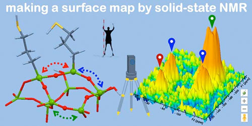
Advanced NMR captures new details in nanoparticle structures(Nanowerk News) Advanced nuclear magnetic resonance (NMR) techniques at the U.S. Department of Energy’s Ames Laboratory have revealed surprising details about the structure of a key group of materials in nanotechnology, mesoporous silica nanoparticles (MSNs), and the placement of their active chemical sites (ACS Catalysis, "Spatial distribution of silica-bound catalytic organic functional groups can now be revealed by conventional and DNP-enhanced solid-state NMR methods"). |
|
| MSNs are honeycombed with tiny (about 2-15 nm wide) three-dimensionally ordered tunnels or pores, and serve as supports for organic functional groups tailored to a wide range of needs. With possible applications in catalysis, chemical separations, biosensing, and drug delivery, MSNs are the focus of intense scientific research. | |
 |
|
| “Since the development of MSNs, people have been trying to control the way they function,” said Takeshi Kobayashi, an NMR scientist with the Division of Chemical and Biological Sciences at Ames Laboratory. “Research has explored doing this through modifying particle size and shape, pore size, and by deploying various organic functional groups on their surfaces to accomplish the desired chemical tasks. However, understanding of the results of these synthetic efforts can be very challenging.” | |
| Ames Laboratory scientist Marek Pruski explained that despite the existence of different techniques for MSNs’ functionalization, no one knew exactly how they were different. In particular, atomic-scale description of how the organic groups were distributed on the surface was lacking until recently. | |
| “It is one thing to detect and quantify these functional groups, or even determine their structure,” said Pruski. “But elucidating their spatial arrangement poses additional challenges. Do they reside on the surfaces or are they partly embedded in the silica walls? Are they uniformly distributed on surfaces? If there are multiple types of functionalities, are they randomly mixed or do they form domains? Conventional NMR, as well as other analytical techniques, have struggled to provide satisfactory answers to these important questions.” | |
| Kobayashi, Pruski, and other researchers used DNP-NMR to obtain a much clearer picture of the structures of functionalized MSNs. “DNP” stands for “dynamic nuclear polarization,” a method which uses microwaves to excite unpaired electrons in radicals and transfer their high spin polarization to the nuclei in the sample being analyzed, offering drastically higher sensitivity, often by two orders of magnitude, and even larger savings of experimental time. | |
| Conventional NMR, which measures the responses of the nuclei of atoms placed in a magnetic field to direct radio-frequency excitation, lacks the sensitivity needed to identify the internuclear interactions between different sites and functionalities on surfaces. When paired with DNP, as well as fast magic angle spinning (MAS), NMR can be used to detect such interactions with unprecedented sensitivity. | |
| Not only did the DNP-NMR methods elicit the atomic-scale location and distribution of the functional groups, but the results disproved some of the existing notions of how MSNs are made and how the different synthetic strategies influenced the dispersion of functional groups throughout the silica pores. | |
| “By examining the role of various experimental conditions, our NMR techniques can give scientists the mechanistic insight they need to guide the synthesis of MSNs in a more controlled way,” said Kobayashi. |
| Source: Ames Laboratory | |
|
Subscribe to a free copy of one of our daily Nanowerk Newsletter Email Digests with a compilation of all of the day's news. |
