Extracellular Matrix: Definition, Structure, Function, and Biomedical Uses
What Is the Extracellular Matrix?
Definition: The extracellular matrix is the non-cellular network of proteins and polysaccharides that surrounds cells, gives tissues mechanical structure, and provides biochemical and mechanical signals that regulate cell behavior.
The extracellular matrix (ECM) is the non-cellular, three-dimensional network of molecules secreted by cells that surrounds and supports them within virtually every tissue and organ. It is not inert packing material: the matrix provides physical scaffolding that holds cells in place and gives tissues their shape and mechanical strength, and it also delivers a continuous stream of biochemical and mechanical signals that guide how cells grow, move, specialize and die. In molecular biology the ECM is understood as an active participant in tissue behavior rather than a passive backdrop.
At a glance:
- Extracellular matrix: the non-cellular network surrounding cells in all tissues
- Main molecular components: collagens and elastin, adhesive glycoproteins, proteoglycans and glycosaminoglycans
- Most abundant protein: collagen (~30% of all body protein)
- Two organizational forms: basement membrane and interstitial matrix
- Full inventory: the matrisome, ~300 core proteins in mammals
- Key biomedical use: scaffolds for tissue engineering
Although the matrix is fundamentally built from water, proteins and polysaccharides, no two tissues share the same matrix. Each tissue assembles a matrix with a distinctive composition and architecture, generated during development through a continuous two-way dialogue between the resident cells and the surrounding matrix they build. In some tissues the ECM dominates the structure: it accounts for the great majority of the dry mass of bone, cartilage and tendon, where relatively few cells maintain a vast scaffold that bears mechanical load.
The matrix exists in two broad organizational forms. The basement membrane is a thin, dense sheet that underlies sheets of epithelial and endothelial cells and separates them from the tissue beneath. The interstitial matrix is the looser, more porous network that fills the spaces between cells in connective tissue. The importance of these structures is underscored by the wide range of inherited disorders, from mild to life-threatening, that arise when a single matrix protein is defective.
Composition of the Extracellular Matrix
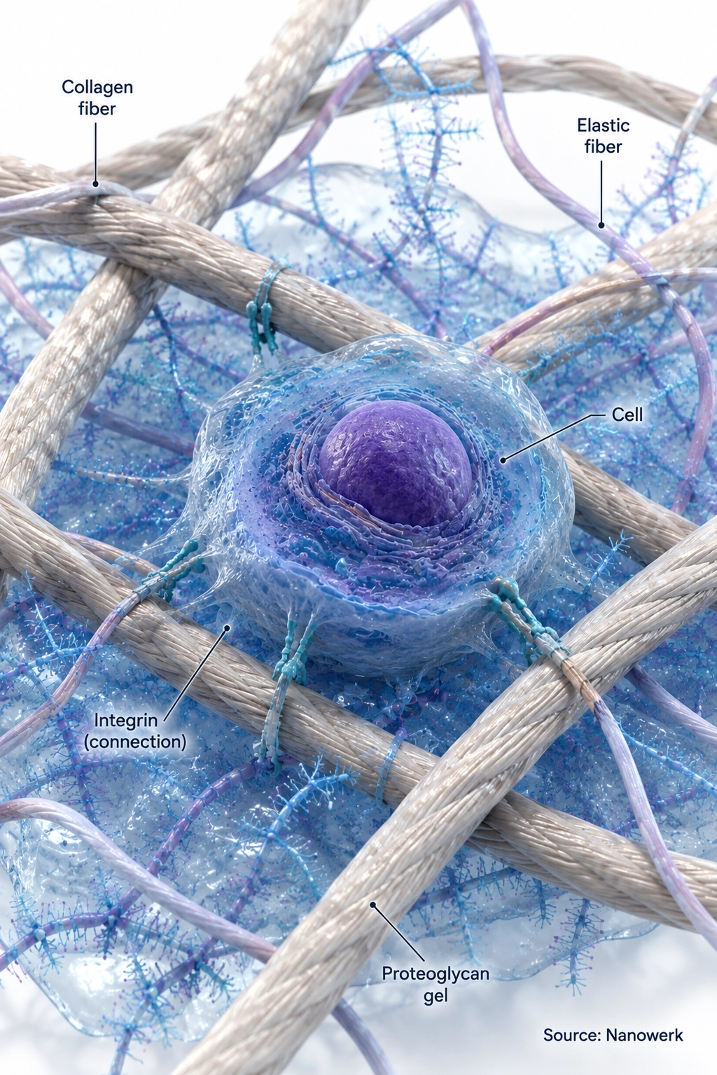
The matrix is built from three broad classes of macromolecule embedded in a hydrated gel. The first class is the fibrous structural proteins, dominated by the collagens, which form ropes and sheets that give tissues tensile strength, together with elastin, which lets skin, lung and blood vessels stretch and recoil. Collagen is the single most abundant protein in mammals, making up on the order of 30 percent of total body protein, and at least 28 distinct collagen types have been described in humans.
The second class is the glycoproteins, which act mainly as adhesive and organizing molecules. Fibronectin links cells to the surrounding fibrillar network and helps direct cell migration during wound healing and development, while laminin is a defining structural component of the basement membrane. The third class is the proteoglycans, each consisting of a core protein decorated with long, negatively charged sugar chains called glycosaminoglycans. These molecules trap large amounts of water, resist compression, and create the cushioning gel that fills the interstitial space. Hyaluronan is an unusually large glycosaminoglycan that is not attached to a core protein, but it contributes strongly to this water-holding capacity.
These classes are not independent. Together, collagen fibers, elastic fibers, adhesive glycoproteins and water-swollen proteoglycans form an integrated material. Its mechanical and signaling properties come from the way these components are combined, cross-linked and remodeled. The table below summarizes the principal component classes and their roles.
| Component class | Representative members | Primary role in the matrix |
|---|---|---|
| Fibrous structural proteins | Collagens (types I, II, III, IV), elastin | Tensile strength, load bearing, elasticity and recoil |
| Adhesive glycoproteins | Fibronectin, laminin, tenascin | Cell attachment, matrix organization, guidance of cell migration |
| Proteoglycans and glycosaminoglycans | Aggrecan, perlecan, hyaluronan, heparan sulfate | Resistance to compression, hydration, growth-factor binding |
| Matrisome-associated remodeling proteins | Matrix metalloproteinases, lysyl oxidase | Degradation, cross-linking and continuous remodeling |
A non-obvious point in this comparison is that the matrix-modifying enzymes are part of what defines the matrix as a living, changing structure rather than a fixed scaffold. The same collagen network that gives a tendon its strength is continuously cut, replaced and re-cross-linked, so the matrix at any moment reflects an ongoing balance between synthesis and breakdown rather than a finished product laid down once.
The Matrisome: A Complete Inventory of Matrix Proteins
Because many matrix proteins share recognizable structural modules and secretion features, researchers can computationally predict much of the ECM-encoding gene set from genome data. This complete inventory is called the matrisome. It is divided into a core matrisome of roughly 300 proteins in mammals, comprising the collagens, proteoglycans and glycoproteins, plus a larger group of matrisome-associated proteins, including the enzymes that remodel the matrix, secreted signaling factors that bind to it, and other ECM-binding proteins.
The matrisome concept, defined computationally and then validated experimentally using proteomics and mass spectrometry, gave the field a shared parts list. It allows researchers to ask which of those several hundred proteins are present in a given organ, how the mixture shifts in disease, and which components change first during processes such as tumor progression. Curated matrisome databases now underpin much of modern ECM research and make it possible to compare the matrix composition of different tissues and tumors on a common footing.
Basement Membrane Versus Interstitial Matrix
The basement membrane is a specialized sheet roughly 50 to 100 nanometers thick that lies beneath every epithelium and surrounds muscle fibers, fat cells and peripheral nerves. Its scaffold is built principally from type IV collagen and laminin, organized into two interlocking networks that are bridged by the proteoglycan perlecan and the glycoprotein nidogen. This thin layer acts as a selective barrier, a polarity cue that tells epithelial cells which side faces outward, and a track that guides cell movement during development and repair.
The interstitial matrix, by contrast, is the bulky three-dimensional network of connective tissue. It is dominated by fibrillar type I collagen, reinforced with fibronectin and embedded in a proteoglycan gel, and it determines the bulk stiffness and resilience of organs such as skin, tendon and the walls of blood vessels. Specialized variants extend this basic plan: the calcified collagen of bone, the compression-resistant aggrecan-rich matrix of cartilage, and the proteoglycan-rich perineuronal nets that stabilize connections in the brain are all tissue-specific elaborations of the same molecular toolkit.
How the Extracellular Matrix Functions
The matrix does far more than hold cells together. Its functions begin with assembly: cells secrete soluble precursors that are processed, cross-linked and organized outside the cell into insoluble fibers and sheets, with enzymes such as lysyl oxidase forming the covalent bonds that lock collagen and elastin into a durable network. The architecture that results is not random; fiber orientation, pore size and stiffness are tuned to the mechanical demands of each tissue.
Cells read the matrix through specialized surface receptors, the most important of which are the integrins. Integrins recognize short binding motifs in matrix proteins, the best known being the arginine-glycine-aspartate (RGD) sequence in fibronectin, also presented by short synthetic peptides used to make biomaterials cell-adhesive. When an integrin engages the matrix it nucleates a cluster of intracellular proteins that links the matrix to the cell's internal skeleton and launches biochemical cascades, a form of signal transduction that controls survival, division and movement.
A central feature of these connections is that they carry mechanical as well as chemical information. Through a process called mechanotransduction, cells physically pull on the matrix and convert its resistance, a soft brain-like gel versus a stiff bone-like substrate, into changes in gene expression and fate. Matrix stiffness alone can strongly bias stem-cell fate in controlled culture systems, which is why matrix mechanics has become a core consideration in cell culture and biomaterial design.
The matrix also serves as a reservoir. Growth factors and other signaling molecules bind to proteoglycans and glycoproteins and are held in place until released, for example by enzymatic cleavage during tissue repair. This lets the matrix store and present signals locally and on demand. All of these functions depend on continuous remodeling: matrix metalloproteinases and related enzymes degrade specific components, freeing space for cells to migrate, releasing stored factors, and generating bioactive protein fragments that themselves act as signals during development, wound healing and disease.
Role in Disease
Because the matrix governs tissue architecture and cell behavior, its disruption is central to many diseases. Fibrosis is the prototypical matrix disease: in response to chronic injury, cells deposit excess collagen and other components faster than they can be removed, progressively stiffening and scarring tissue. This drives organ failure in conditions such as liver cirrhosis, idiopathic pulmonary fibrosis and chronic kidney disease, where the accumulating matrix physically displaces functional tissue.
In cancer, the matrix surrounding a tumor is frequently denser, stiffer and compositionally abnormal compared with healthy tissue. This altered matrix can promote tumor cell survival, distort normal signaling, and create physical paths and mechanical cues that help malignant cells invade and spread to distant sites. The disease-specific changes in matrix proteins are increasingly studied as a source of diagnostic biomarkers and as targets for therapies aimed at the tumor's surroundings rather than the cancer cells alone.
Inherited defects in matrix components cause a distinct group of disorders. Mutations in a collagen gene can produce osteogenesis imperfecta, in which bones become brittle, while defects in fibrillin disrupt elastic fibers in Marfan syndrome, and several forms of Ehlers-Danlos syndrome stem from faulty collagen processing. Defects in basement-membrane components can also disrupt filtration barriers in the kidney or tissue integrity in skin and muscle. Gradual matrix changes also accompany normal aging, as accumulated cross-linking stiffens tissues and slows the repair processes that depend on a healthy, remodelable matrix.
Applications in Tissue Engineering and Regenerative Medicine
The matrix is a central tool in tissue engineering precisely because cells respond so strongly to the scaffold around them. One major strategy is decellularization: an organ or tissue is stripped of its cells with detergents, leaving behind the natural matrix with its architecture and biochemical cues intact. The resulting scaffold can be reseeded with a patient's own cells, an approach being pursued for skin, heart valves, blood vessels and more complex organs in regenerative medicine.
Matrix-derived materials are also widely used in the laboratory. Reconstituted basement-membrane preparations and purified collagen gels provide the soft, signal-rich environment that allows stem cells to grow into three-dimensional organoids that mimic real tissues, and they form the biological component of many organ-on-a-chip devices. In 3D bioprinting, matrix proteins such as collagen and gelatin, sometimes combined with decellularized tissue, are formulated into printable bioinks that hold cells in a defined three-dimensional pattern.
A complementary direction is the design of synthetic and engineered matrices that reproduce only the cues that matter. Using protein engineering and defined chemistries, researchers build hydrogels with tunable stiffness, controlled degradation and grafted adhesion peptides, giving precise control over the signals a cell receives. The broad goal across biotechnology is to move from using whatever matrix nature provides toward building matrices to specification, normalizing a diseased matrix or instructing cells to regenerate tissue.
Frequently Asked Questions
What is the extracellular matrix made of? The extracellular matrix is made of water, fibrous structural proteins such as collagens and elastin, glycoproteins such as fibronectin and laminin, and proteoglycans built from a core protein decorated with long glycosaminoglycan sugar chains. Collagen is the most abundant protein, accounting for roughly 30 percent of all protein in the mammalian body. The exact mixture and architecture differ from tissue to tissue, which is why bone, cartilage, tendon and brain each have a distinct matrix.
What is the difference between the basement membrane and the interstitial matrix? The basement membrane is a thin, sheet-like matrix built mainly from type IV collagen and laminin that underlies epithelial and endothelial cell layers and separates them from the underlying tissue. The interstitial matrix is the looser three-dimensional network that fills the space between cells in connective tissue and is rich in fibrillar type I collagen, fibronectin and proteoglycans. The two forms differ in composition, architecture and mechanical role.
What is the matrisome? The matrisome is the complete set of genes and proteins that make up the extracellular matrix and the factors that associate with it. It is divided into a core matrisome of roughly 300 proteins in mammals, comprising collagens, proteoglycans and glycoproteins, plus a larger group of matrisome-associated proteins such as ECM-remodeling enzymes, secreted factors and ECM-binding proteins. The concept was defined computationally and validated by proteomics.
How does the extracellular matrix communicate with cells? Cells attach to the matrix through surface receptors, most importantly the integrins, which bind specific motifs in matrix proteins such as the RGD sequence in fibronectin. These attachments transmit both chemical signals and mechanical force across the cell membrane, a process called mechanotransduction, allowing cells to sense matrix stiffness and composition and adjust their growth, shape, movement and gene expression accordingly.
Why is the extracellular matrix important in cancer and fibrosis? In fibrosis, excess collagen and other matrix components accumulate and stiffen tissue, impairing organ function in conditions such as liver cirrhosis and pulmonary fibrosis. In cancer, the matrix surrounding a tumor is often abnormally dense and stiff, which can promote tumor cell survival, invasion and spread, and altered matrix proteins are being explored as biomarkers and drug targets.
Further Reading
Journal of Cell Science, The Extracellular Matrix at a Glance
Molecular & Cellular Proteomics, The Matrisome: In Silico Definition and In Vivo Characterization by Proteomics of Normal and Tumor Extracellular Matrices
Nature Reviews Molecular Cell Biology, Mechanisms of Assembly and Remodelling of the Extracellular Matrix
Nature Reviews Cancer, The Matrix in Cancer
